
Ethmoidal air cells
Citation, DOI, disclosures and article data
At the time the article was created Maxime St-Amant had no recorded disclosures.
View Maxime St-Amant's current disclosuresAt the time the article was last revised Tariq Walizai had no financial relationships to ineligible companies to disclose.
View Tariq Walizai's current disclosures- Ethmoidal air cell
- Ethmoidal sinuses
- Ethmoid sinuses
- Ethmoid air cells
- Ethmoid air cell
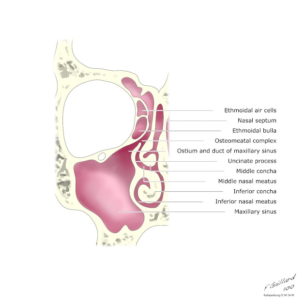
The ethmoidal air cells, also known less commonly as the ethmoidal sinuses, form one of the four pairs of paranasal sinuses. They are located within the single, midline ethmoid bone.
On this page:
Summary
location: between the orbit and the nasal cavity, within the ethmoid labyrinth of the ethmoid bone
blood supply: supraorbital, anterior and posterior ethmoidal and sphenopalatine arteries
innervation: anterior and posterior ethmoidal and supraorbital nerves
Gross anatomy
A collection of air cells (3-18 in number) separated by bony septa within each side of the lateral mass, or labyrinth, of the ethmoid bone.
They are separated into anterior and posterior groups by the basal lamella, the lateral attachment of the middle turbinate to the lamina papyracea. Historically the ethmoid sinuses were subdivided into 3 groups of air cells: the anterior, middle and posterior ethmoidal air cells. The middle group are now incorporated into the anterior group.
The anterior ethmoidal air cells drain to the hiatus semilunaris and middle meatus via the ethmoid bulla, which forms parts of the ostiomeatal complex. The posterior ethmoidal air cells drain to the superior meatus via the sphenoethmoidal recess 2.
Some of the ethmoidal air cells have been given specific names, because of their importance in surgical procedures or involvement in head and neck pathologies:
frontal recess cells, including the agger nasi cells
Arterial supply
From the ophthalmic branch of the internal carotid artery, the supraorbital, anterior and posterior ethmoidal arteries supply the ethmoid air cells with the sphenopalatine artery (a branch of the maxillary artery) also contributing. Thus, the ethmoid air cells are supplied by branches of both the internal and external carotid arteries.
Lymphatic drainage
Lymph from the ethmoid air cells drains to the submandibular and retropharyngeal group of nodes.
Innervation
The posterior ethmoidal air cells, along with the sphenoid sinus, are supplied by the posterior ethmoidal nerve, whereas the anterior ethmoidal nerve supplies the anterior ethmoidal air cells. Both these nerves are extraconal branches of the nasociliary nerve, a branch of the ophthalmic division of the trigeminal nerve.
Development
They are present at birth, and they develop rapidly from 0-4 years of age. They further mature from 8-12 years of age through puberty.
References
- 1. Jones N, Strobl A, Holland I. A Study of the CT Findings in 100 Patients with Rhinosinusitis and 100 Controls. Clin Otolaryngol Allied Sci. 1997;22(1):47-51. doi:10.1046/j.1365-2273.1997.00862.x - Pubmed
- 2. H. Ric Harnsberger, André J. Macdonald. Diagnostic and Surgical Imaging Anatomy. (2006) ISBN: 9781931884297 - Google Books
- 3. Keith L. Moore, Arthur F. Dalley, A. M. R. Agur. Clinically Oriented Anatomy. (2013) ISBN: 9781451119459 - Google Books
- 4. Last, R. J., McMinn, R. M. H.. Last's Anatomy, Regional and Applied. (1994) ISBN: 044304662X - Google Books
- 5. Paul Butler, Adam Mitchell, Jeremiah C. Healy et al. Applied Radiological Anatomy. (2012) ISBN: 9780521766661 - Google Books
- 6. Robert H. Whitaker, Neil R. Borley. Instant Anatomy. (2000) ISBN: 9780632054039 - Google Books
Incoming Links
- Middle meatus
- Patterns of sinonasal obstruction
- Transsphenoidal basilar skull fracture
- Supreme meatus
- Nasal bone
- Hiatus semilunaris
- Orbital nerve supply
- Sinonasal adenocarcinoma
- Anterior ethmoidal artery
- Lamina papyracea
- Paranasal sinus mucocele
- Basal lamella
- Allergic fungal sinusitis
- Ossifying fibromyxoid tumour
- Superior meatus
- Anterior ethmoidal notch
- Pharyngeal nerve
- Sinonasal undifferentiated carcinoma
- Olfactory neuroblastoma
- Ethmoid bulla
- Ethmoid mucocele
- Paranasal sinus development (Gray's illustration)
- Esthesioneuroblastoma
- Sinonasal angiomatous polyp
- Orbital medial wall blow-out fracture and retrobulbar emphysema
- Fronto-ethmoidal sinus mucocele
- Osteoma of the ethmoid air cell
- Orbital medial wall and floor blow-out fracture
- Orbital medial wall blow-out fracture
- Dacryocystitis and dacryocystocele
- Blow-out fracture of the orbit and retrobulbar hemorrhage
- Subperiosteal abscess of the orbit
- Orbital medial wall blow-out fracture
- Frontoethmoidal mucocele
- Large frontal osteoma
- Ocular globe rupture and orbital blow-out fracture
- Orbital blow-out fracture and extraconal hematoma
- Orbital Blow-out fracture and retrobulbar hemorrhage
- Orbital Blow-out fracture and inferior rectus muscle transection
- Ethmoid sinus osteoma
Related articles: Anatomy: Head and neck
- skeleton of the head and neck
-
cranial vault
- scalp (mnemonic)
- fontanelle
-
sutures
- calvarial
- facial
- frontozygomatic suture
- frontomaxillary suture
- frontolacrimal suture
- frontonasal suture
- temporozygomatic suture
- zygomaticomaxillary suture
- parietotemporal suture (parietomastoid suture)
- occipitotemporal suture (occipitomastoid suture)
- sphenofrontal suture
- sphenozygomatic suture
- spheno-occipital suture (not a true suture)
- lacrimomaxillary suture
- nasomaxillary suture
- internasal suture
- basal/internal
- skull landmarks
- frontal bone
- temporal bone
- parietal bone
- occipital bone
- skull base (foramina)
-
facial bones
- midline single bones
- paired bilateral bones
- cervical spine
- hyoid bone
- laryngeal cartilages
-
cranial vault
- muscles of the head and neck
- muscles of the tongue (mnemonic)
- muscles of mastication
-
facial muscles
- epicranius muscle
- circumorbital and palpebral muscles
- nasal muscles
-
buccolabial muscles
- elevators, retractors and evertors of the upper lip
- levator labii superioris alaeque nasalis muscle
- levator labii superioris muscle
- zygomaticus major muscle
- zygomaticus minor muscle
- levator anguli oris muscle
- malaris muscle
- risorius muscle
- depressors, retractors and evertors of the lower lip
- depressor labii inferioris muscle
- depressor anguli oris muscle
- mentalis muscle
- compound sphincter
-
orbicularis oris muscle
- incisivus labii superioris muscle
- incisivus labii inferioris muscle
-
orbicularis oris muscle
- muscle of mastication
- modiolus
- elevators, retractors and evertors of the upper lip
- muscles of the middle ear
- orbital muscles
- muscles of the soft palate
- pharyngeal muscles
- suprahyoid muscles
- infrahyoid muscles
- intrinsic muscles of the larynx
- muscles of the neck
- platysma muscle
- longus colli muscle
- longus capitis muscle
- scalenus anterior muscle
- scalenus medius muscle
- scalenus posterior muscle
- scalenus pleuralis muscle
- sternocleidomastoid muscle
-
suboccipital muscles
- rectus capitis posterior major muscle
- rectus capitis posterior minor muscle
- obliquus capitis superior muscle
- obliquus capitis inferior muscle
- accessory muscles of the neck
- deep cervical fascia
-
deep spaces of the neck
- anterior cervical space
- buccal space
- carotid space
- danger space
- deep cervical fascia
- infratemporal fossa
- masticator space
- parapharyngeal space
- stylomandibular tunnel
- parotid space
- pharyngeal (superficial) mucosal space
- perivertebral space
- posterior cervical space
- pterygopalatine fossa
- retropharyngeal space
- suprasternal space (of Burns)
- visceral space
- surgical triangles of the neck
- orbit
- ear
- paranasal sinuses
- upper respiratory tract
- viscera of the neck
- blood supply of the head and neck
-
arterial supply
-
common carotid artery
- carotid body
- carotid bifurcation
- subclavian artery
- variants
-
common carotid artery
- venous drainage
-
arterial supply
- innervation of the head and neck
-
cranial nerves
- olfactory nerve (CN I)
- optic nerve (CN II)
- oculomotor nerve (CN III)
- trochlear nerve (CN IV)
-
trigeminal nerve (CN V) (mnemonic)
- trigeminal ganglion
- ophthalmic division
- maxillary division
- mandibular division
- abducens nerve (CN VI)
- facial nerve (CN VII)
-
vestibulocochlear nerve (CN VIII)
- vestibular ganglion (Scarpa's ganglion)
- glossopharyngeal nerve (CN IX)
- vagus nerve (CN X)
- (spinal) accessory nerve (CN XI)
- hypoglossal nerve (CN XII)
- parasympathetic ganglia of the head and neck
- cervical sympathetic ganglia
- greater occipital nerve
- third occipital nerve
-
cervical plexus
- muscular branches
- longus capitis
- longus colli
- scalenes
- geniohyoid
- thyrohyoid
-
ansa cervicalis
- omohyoid (superior and inferior bellies separately)
- sternothyroid
- sternohyoid
- phrenic nerve
- contribution to the accessory nerve (CN XI)
- cutaneous branches
- muscular branches
- brachial plexus
- pharyngeal plexus
-
cranial nerves
- lymphatic drainage of the head and neck
- embryological development of the head and neck


 Unable to process the form. Check for errors and try again.