
Dual-energy CT has a number of clinical applications in neuroimaging particularly in the realm of material composition and virtual non-contrast imaging.
On this page:
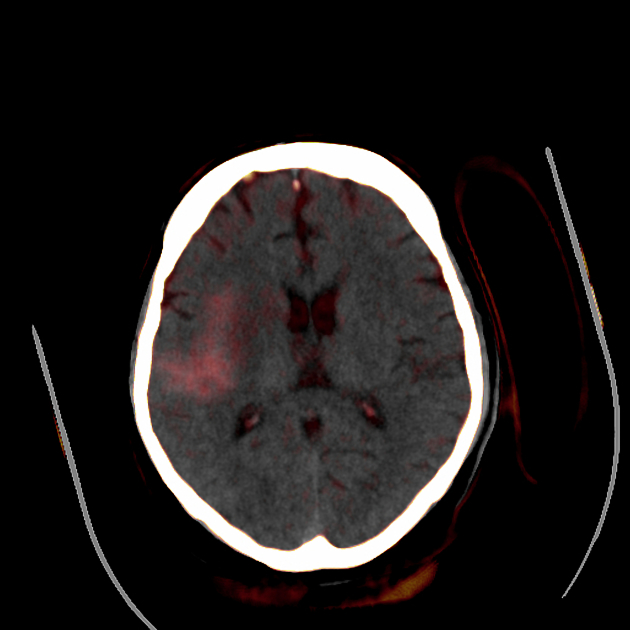
Differentiation of hemorrhage from iodinated contrast
Contrast staining of the brain parenchyma post iodinated contrast can lead to interpretation issues, especially when the density of contrast is similar to that of blood. Virtual non-contrast imaging is an accurate 1 method in differentiating intracranial hemorrhage from iodinated contrast staining following interventional procedures such as endovascular clot retrieval. Similarly, differentiation can be made between enhancing subdural hygroma (effusion) and subdural hematoma 5.
Quantification of iodine leak in traumatic hemorrhagic contusions
Using iodine quantification one can measure the amount of iodine leak within a core and the penumbra of hemorrhagic contusions after contrast-enhanced whole-body CT 2.
Bone removal
Conventionally removing bone in was either a manual process or a threshold related algorithm (that often subsequently involves a manual process). Using three material differentiation, dual-energy CT scans can be post-processed to remove bone which is particularly advantageous in both angiography and venography 3. It is not perfect and has been found to overestimate stenosis when there is calcification close to the skull, additionally the presence of blooming artifact caused by calcifications can lead to artifacts in the post-processing too 3.
Optimizing imaging
Using monoenergetic image reconstruction one can change the energy levels depending on the region of interest, for example using 75 keV at the posterior fossa to reduce beam hardening artifact 4.


 Unable to process the form. Check for errors and try again.