
Otic ganglion
Citation, DOI, disclosures and article data
At the time the article was created Prashant Mudgal had no recorded disclosures.
View Prashant Mudgal's current disclosuresAt the time the article was last revised Roland Zhang had no financial relationships to ineligible companies to disclose.
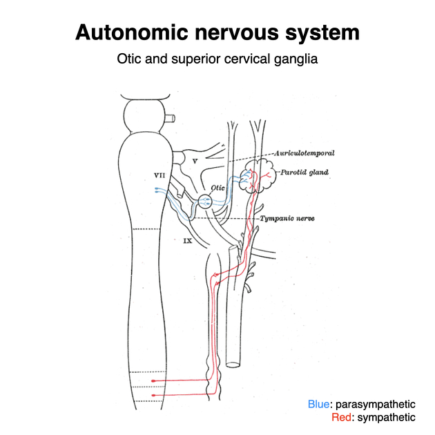
View Roland Zhang's current disclosuresThe otic ganglion is one of four parasympathetic ganglia of the head and neck. It receives parasympathetic fibers from the glossopharyngeal nerve. It supplies the parotid gland with not just parasympathetic, but also sympathetic and sensory fibers that pass through the gland.
On this page:
Images:
Gross anatomy
small and disc shaped ganglion
located in the infratemporal fossa
it lies immediately below the foramen ovale, medial to the mandibular division of the trigeminal nerve
supplies secretomotor fibers to the parotid gland
Roots
-
parasympathetic root
from the inferior salivary nucleus via the glossopharyngeal nerve and branch of the tympanic nerve (Jacobson's nerve) to form the tympanic plexus in the middle ear and then exit as the lesser petrosal nerve
the lesser petrosal nerve then passes from the foramen ovale to the ganglion and then fibers reach the parotid gland as the auriculotemporal nerve
-
sympathetic root
via the middle meningeal artery, vasomotor fibers from the superior cervical ganglion pass through the ganglion without synapsing and course though the auriculotemporal nerve and supply the parotid gland
-
sensory root
via the auriculotemporal nerve to the parotid gland
Branches
Related pathology
Frey syndrome: where salivary stimuli induces perspiration and erythema at the parotid
References
- 1. Shimizu T. Distribution and pathway of the cerebrovascular nerve fibers from the otic ganglion in the rat: anterograde tracing study. J. Auton. Nerv. Syst. 1994;49 (1): 47-54. Pubmed citation
- 2. Kahle W, Frotscher M. Color Atlas and Textbook of Human Anatomy: Nervous system and sensory organs. Thieme. (2003) ISBN:1588900649. Read it at Google Books - Find it at Amazon
- 3. Last's anatomy, regional and applied. Churchill Livingstone. ISBN:044304662X. Read it at Google Books - Find it at Amazon
- 4. Moore KL, Agur AMR, Dalley AF. Clinically oriented anatomy. LWW. ISBN:1451119453. Read it at Google Books - Find it at Amazon
Incoming Links
- Parasympathetic nervous system
- Lesser petrosal nerve
- Pineal gland
- Middle ear
- Autonomic ganglia and plexuses
- Infratemporal fossa
- Inferior salivatory nucleus
- Auriculotemporal nerve
- Facial nerve
- Jacobson nerve
- Parasympathetic ganglia in the head and neck
- Glossopharyngeal nerve
- Foramen ovale (skull)
- Foramen ovale contents (mnemonic)
- Buccal nerve
- Inferior alveolar nerve
- Parotid gland
- Nerve to medial pterygoid
Related articles: Anatomy: Head and neck
- skeleton of the head and neck
-
cranial vault
- scalp (mnemonic)
- fontanelle
-
sutures
- calvarial
- facial
- frontozygomatic suture
- frontomaxillary suture
- frontolacrimal suture
- frontonasal suture
- temporozygomatic suture
- zygomaticomaxillary suture
- parietotemporal suture (parietomastoid suture)
- occipitotemporal suture (occipitomastoid suture)
- sphenofrontal suture
- sphenozygomatic suture
- spheno-occipital suture (not a true suture)
- lacrimomaxillary suture
- nasomaxillary suture
- internasal suture
- basal/internal
- skull landmarks
- frontal bone
- temporal bone
- parietal bone
- occipital bone
- skull base (foramina)
-
facial bones
- midline single bones
- paired bilateral bones
- cervical spine
- hyoid bone
- laryngeal cartilages
-
cranial vault
- muscles of the head and neck
- muscles of the tongue (mnemonic)
- muscles of mastication
-
facial muscles
- epicranius muscle
- circumorbital and palpebral muscles
- nasal muscles
-
buccolabial muscles
- elevators, retractors and evertors of the upper lip
- levator labii superioris alaeque nasalis muscle
- levator labii superioris muscle
- zygomaticus major muscle
- zygomaticus minor muscle
- levator anguli oris muscle
- malaris muscle
- risorius muscle
- depressors, retractors and evertors of the lower lip
- depressor labii inferioris muscle
- depressor anguli oris muscle
- mentalis muscle
- compound sphincter
-
orbicularis oris muscle
- incisivus labii superioris muscle
- incisivus labii inferioris muscle
-
orbicularis oris muscle
- muscle of mastication
- modiolus
- elevators, retractors and evertors of the upper lip
- muscles of the middle ear
- orbital muscles
- muscles of the soft palate
- pharyngeal muscles
- suprahyoid muscles
- infrahyoid muscles
- intrinsic muscles of the larynx
- muscles of the neck
- platysma muscle
- longus colli muscle
- longus capitis muscle
- scalenus anterior muscle
- scalenus medius muscle
- scalenus posterior muscle
- scalenus pleuralis muscle
- sternocleidomastoid muscle
-
suboccipital muscles
- rectus capitis posterior major muscle
- rectus capitis posterior minor muscle
- obliquus capitis superior muscle
- obliquus capitis inferior muscle
- accessory muscles of the neck
- deep cervical fascia
-
deep spaces of the neck
- anterior cervical space
- buccal space
- carotid space
- danger space
- deep cervical fascia
- infratemporal fossa
- masticator space
- parapharyngeal space
- stylomandibular tunnel
- parotid space
- pharyngeal (superficial) mucosal space
- perivertebral space
- posterior cervical space
- pterygopalatine fossa
- retropharyngeal space
- suprasternal space (of Burns)
- visceral space
- surgical triangles of the neck
- orbit
- ear
- paranasal sinuses
- upper respiratory tract
- viscera of the neck
- blood supply of the head and neck
-
arterial supply
-
common carotid artery
- carotid body
- carotid bifurcation
- subclavian artery
- variants
-
common carotid artery
- venous drainage
-
arterial supply
- innervation of the head and neck
-
cranial nerves
- olfactory nerve (CN I)
- optic nerve (CN II)
- oculomotor nerve (CN III)
- trochlear nerve (CN IV)
-
trigeminal nerve (CN V) (mnemonic)
- trigeminal ganglion
- ophthalmic division
- maxillary division
- mandibular division
- abducens nerve (CN VI)
- facial nerve (CN VII)
-
vestibulocochlear nerve (CN VIII)
- vestibular ganglion (Scarpa's ganglion)
- glossopharyngeal nerve (CN IX)
- vagus nerve (CN X)
- (spinal) accessory nerve (CN XI)
- hypoglossal nerve (CN XII)
- parasympathetic ganglia of the head and neck
- cervical sympathetic ganglia
- greater occipital nerve
- third occipital nerve
-
cervical plexus
- muscular branches
- longus capitis
- longus colli
- scalenes
- geniohyoid
- thyrohyoid
-
ansa cervicalis
- omohyoid (superior and inferior bellies separately)
- sternothyroid
- sternohyoid
- phrenic nerve
- contribution to the accessory nerve (CN XI)
- cutaneous branches
- muscular branches
- brachial plexus
- pharyngeal plexus
-
cranial nerves
- lymphatic drainage of the head and neck
- embryological development of the head and neck
Related articles: Anatomy: General
- anatomic position
-
anatomic nomenclature
-
Terminologia Anatomica
- superseded nomenclature
-
Terminologia Anatomica
- anatomic variants
- labeled imaging anatomy cases
- regional anatomy
- systems anatomy
- endocrine system
- lymphatic system
- reticuloendothelial system
- nervous system
- systems based on location
- systems based on function
- somatic nervous system
-
autonomic nervous system
- sympathetic nervous system
- parasympathetic nervous system
-
autonomic ganglia and plexuses
- craniofacial
- cervical
- thoracic
- abdominopelvic
- coccygeal
- histology
- osteology
- skeleton
- bones
- macroscopic structure
- microscopic structure
- bone growth
- fetal bone formation
- developmental ossification
- tubulation
- bone types
- nutrient foramen
- joints
- muscles
- organs
- embryology
- skin
- blood vessels


 Unable to process the form. Check for errors and try again.