

Peripheral nerve sheath tumor
Updates to Article Attributes
Peripheral nerve sheath tumours (PNSTs) are a group of primary neurogenic tumours that arise from nerve sheaths outside of the central nervous system. The vast majority are benign, however, malignant transformation is seen particularly in large tumours and those associated with neurofibromatosis type 1 (NF1).
Pathology
Markers
Many peripheral nerve sheath tumours may express somatostatin receptors 6
Classification
Their imaging appearances, demographics, treatment and prognosis vary greatly and these are therefore discussed separately.
Peripheral nerve sheath tumours may be classified as follows:
-
benign
-
malignant
Radiographic features
Imaging of a solitary peripheral nerve sheath tumour, in most cases, cannot reliably distinguish between the different histological subtypes, and a presumptive diagnosis must take into account the patient demographics, including pre-existing conditions (such as neurofibromatosis type 1 or neurofibromatosis type 2), the location and size of the tumour, and evidence of rapid growth. As such these are discussed separately in each of the aforementioned tumours. A number of features are, however, shared by localised neurogenic tumours of peripheral nerves.
MRI
As a group, localised peripheral nerve sheath tumours demonstrate the following features:
fusiform-shaped mass with tapered ends, with nerve seen leading into and out of the mass
denervation changes in muscles supplied by the involved nerve
Differential diagnosis
Considerations include 7,8:
benign and malignant myxoid tumours, e.g. myxoid liposarcoma, myxofibrosarcoma, fibromyxoid sarcoma
-<p><strong>Peripheral nerve sheath tumours (PNSTs)</strong> are a group of primary <a href="/articles/neurogenic-tumours-1">neurogenic tumours</a> that arise from nerve sheaths outside of the central nervous system. The vast majority are benign, however, malignant transformation is seen particularly in large tumours and those associated with <a href="/articles/neurofibromatosis-type-1">neurofibromatosis type 1 (NF1)</a>. </p><h4>Pathology</h4><h5>Markers</h5><p>Many peripheral nerve sheath tumours may express somatostatin receptors <sup>6</sup></p><h4>Classification</h4><p>Their imaging appearances, demographics, treatment and prognosis vary greatly and these are therefore discussed separately.</p><p>Peripheral nerve sheath tumours may be classified as follows:</p><ul>-<li>-<p>benign</p>-<ul>-<li><p><a href="/articles/schwannoma">schwannoma</a></p></li>-<li>-<p><a href="/articles/neurofibroma">neurofibroma</a></p>-<ul>-<li>-<p><a href="/articles/localised-neurofibroma">localised neurofibroma</a></p>-<ul>-<li><p><a href="/articles/localised-cutaneous-neurofibroma">localised cutaneous neurofibroma</a></p></li>-<li><p><a href="/articles/localised-intraneural-neurofibroma">localised intraneural neurofibroma</a></p></li>-</ul>-</li>-<li><p><a href="/articles/diffuse-cutaneous-neurofibroma">diffuse cutaneous neurofibroma</a></p></li>-<li><p><a href="/articles/plexiform-neurofibroma">plexiform neurofibroma</a></p></li>-</ul>-</li>-<li><p><a href="/articles/perineurioma">perineurioma</a></p></li>-<li><p><a href="/articles/hybrid-nerve-sheath-tumour-1">hybrid nerve sheath tumour</a></p></li>-</ul>-</li>-<li>-<p>malignant</p>-<ul><li><p><a href="/articles/malignant-peripheral-nerve-sheath-tumour">malignant peripheral nerve sheath tumour</a></p></li></ul>-</li>-</ul><h4>Radiographic features</h4><p>Imaging of a solitary peripheral nerve sheath tumour, in most cases, cannot reliably distinguish between the different histological subtypes, and a presumptive diagnosis must take into account the patient demographics, including pre-existing conditions (such as <a href="/articles/neurofibromatosis-type-1">neurofibromatosis type 1</a> or <a href="/articles/neurofibromatosis-type-2-3">neurofibromatosis type 2</a>), the location and size of the tumour, and evidence of rapid growth. As such these are discussed separately in each of the aforementioned tumours. A number of features are, however, shared by localised neurogenic tumours of peripheral nerves.</p><h5>MRI</h5><p>As a group, localised peripheral nerve sheath tumours demonstrate the following features:</p><ul>-<li><p>fusiform-shaped mass with tapered ends, with nerve seen leading into and out of the mass</p></li>-<li><p><a href="/articles/split-fat-sign">split-fat sign</a></p></li>-<li><p><a href="/articles/fascicular-sign">fascicular sign</a></p></li>-<li><p><a href="/articles/target-sign-peripheral-nerve-sheath-tumor">target sign</a></p></li>-<li><p><a href="/articles/denervation-changes-in-muscles">denervation changes in muscles</a> supplied by the involved nerve</p></li>-</ul><h4>Differential diagnosis</h4><p>Considerations include <sup>7,8</sup>:</p><ul>-<li><p><a href="/articles/localised-tenosynovial-giant-cell-tumour-1" title="Localised tenosynovial giant cell tumour">tenosynovial giant cell tumour</a></p></li>-<li><p><a href="/articles/angioleiomyoma" title="Angioleiomyoma">angioleiomyoma</a></p></li>-<li><p><a href="/articles/haemangioma" title="Hemangioma">haemangioma</a></p></li>-<li><p>benign and malignant myxoid tumours, e.g. <a href="/articles/myxoid-liposarcoma" title="Myxoid liposarcoma">myxoid liposarcoma</a>, <a href="/articles/myxofibrosarcoma" title="Myxofibrosarcoma">myxofibrosarcoma</a>, <a href="/articles/low-grade-fibromyxoid-sarcoma" title="Low-grade fibromyxoid sarcoma">fibromyxoid sarcoma</a></p></li>- +<p><strong>Peripheral nerve sheath tumours (PNSTs)</strong> are a group of primary <a href="/articles/neurogenic-tumours-1">neurogenic tumours</a> that arise from nerve sheaths outside of the central nervous system. The vast majority are benign, however, malignant transformation is seen particularly in large tumours and those associated with <a href="/articles/neurofibromatosis-type-1">neurofibromatosis type 1 (NF1)</a>. </p><h4>Pathology</h4><h5>Markers</h5><p>Many peripheral nerve sheath tumours may express somatostatin receptors <sup>6</sup></p><h4>Classification</h4><p>Their imaging appearances, demographics, treatment and prognosis vary greatly and these are therefore discussed separately.</p><p>Peripheral nerve sheath tumours may be classified as follows:</p><ul>
- +<li>
- +<p>benign</p>
- +<ul>
- +<li><p><a href="/articles/schwannoma">schwannoma</a></p></li>
- +<li>
- +<p><a href="/articles/neurofibroma">neurofibroma</a></p>
- +<ul>
- +<li>
- +<p><a href="/articles/localised-neurofibroma">localised neurofibroma</a></p>
- +<ul>
- +<li><p><a href="/articles/localised-cutaneous-neurofibroma">localised cutaneous neurofibroma</a></p></li>
- +<li><p><a href="/articles/localised-intraneural-neurofibroma">localised intraneural neurofibroma</a></p></li>
- +</ul>
- +</li>
- +<li><p><a href="/articles/diffuse-cutaneous-neurofibroma">diffuse cutaneous neurofibroma</a></p></li>
- +<li><p><a href="/articles/plexiform-neurofibroma">plexiform neurofibroma</a></p></li>
- +</ul>
- +</li>
- +<li><p><a href="/articles/perineurioma">perineurioma</a></p></li>
- +<li><p><a href="/articles/hybrid-nerve-sheath-tumour-1">hybrid nerve sheath tumour</a></p></li>
- +</ul>
- +</li>
- +<li>
- +<p>malignant</p>
- +<ul><li><p><a href="/articles/malignant-peripheral-nerve-sheath-tumour">malignant peripheral nerve sheath tumour</a></p></li></ul>
- +</li>
- +</ul><h4>Radiographic features</h4><p>Imaging of a solitary peripheral nerve sheath tumour, in most cases, cannot reliably distinguish between the different histological subtypes, and a presumptive diagnosis must take into account the patient demographics, including pre-existing conditions (such as <a href="/articles/neurofibromatosis-type-1">neurofibromatosis type 1</a> or <a href="/articles/neurofibromatosis-type-2-3">neurofibromatosis type 2</a>), the location and size of the tumour, and evidence of rapid growth. As such these are discussed separately in each of the aforementioned tumours. A number of features are, however, shared by localised neurogenic tumours of peripheral nerves.</p><h5>MRI</h5><p>As a group, localised peripheral nerve sheath tumours demonstrate the following features:</p><ul>
- +<li><p>fusiform-shaped mass with tapered ends, with nerve seen leading into and out of the mass</p></li>
- +<li><p><a href="/articles/split-fat-sign">split-fat sign</a></p></li>
- +<li><p><a href="/articles/fascicular-sign">fascicular sign</a></p></li>
- +<li><p><a href="/articles/target-sign-peripheral-nerve-sheath-tumor">target sign</a></p></li>
- +<li><p><a href="/articles/denervation-changes-in-muscles">denervation changes in muscles</a> supplied by the involved nerve</p></li>
- +</ul><h4>Differential diagnosis</h4><p>Considerations include <sup>7,8</sup>:</p><ul>
- +<li><p><a href="/articles/localised-tenosynovial-giant-cell-tumour-1" title="Localised tenosynovial giant cell tumour">tenosynovial giant cell tumour</a></p></li>
- +<li><p><a href="/articles/angioleiomyoma" title="Angioleiomyoma">angioleiomyoma</a></p></li>
- +<li><p><a href="/articles/haemangioma" title="Hemangioma">haemangioma</a></p></li>
- +<li><p>benign and malignant myxoid tumours, e.g. <a href="/articles/myxoid-liposarcoma" title="Myxoid liposarcoma">myxoid liposarcoma</a>, <a href="/articles/myxofibrosarcoma" title="Myxofibrosarcoma">myxofibrosarcoma</a>, <a href="/articles/low-grade-fibromyxoid-sarcoma" title="Low-grade fibromyxoid sarcoma">fibromyxoid sarcoma</a></p></li>
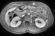
Image 7 MRI (T1 C+ fat sat) ( create )


 Unable to process the form. Check for errors and try again.