
Pituitary region masses include lesions in the sella turcica, suprasellar cistern, parasellar region include cavernous sinuses, and basisphenoid/clivus. Several mnemonics have been popularized, including SATCHMO.
A more comprehensive list includes the following, along with differentiating features:
-
tumors
-
intrasellar
-
pituitary neuroendocrine tumor (PitNET) (commonest in adults)
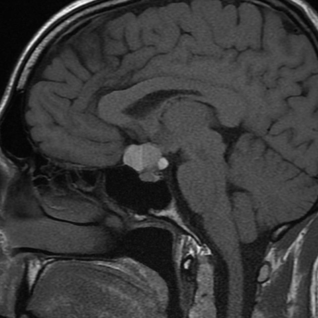
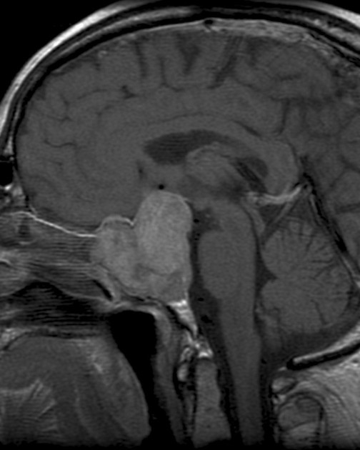
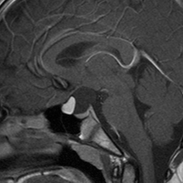
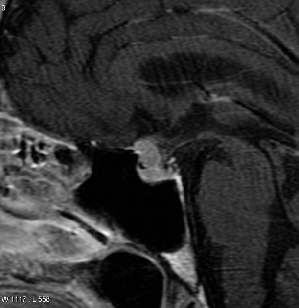
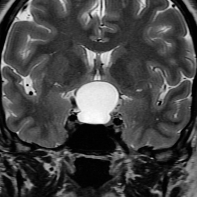
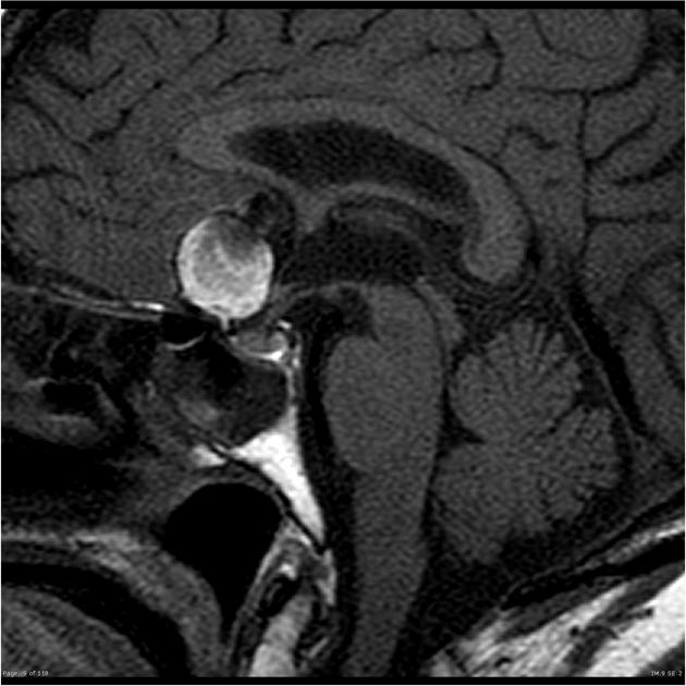
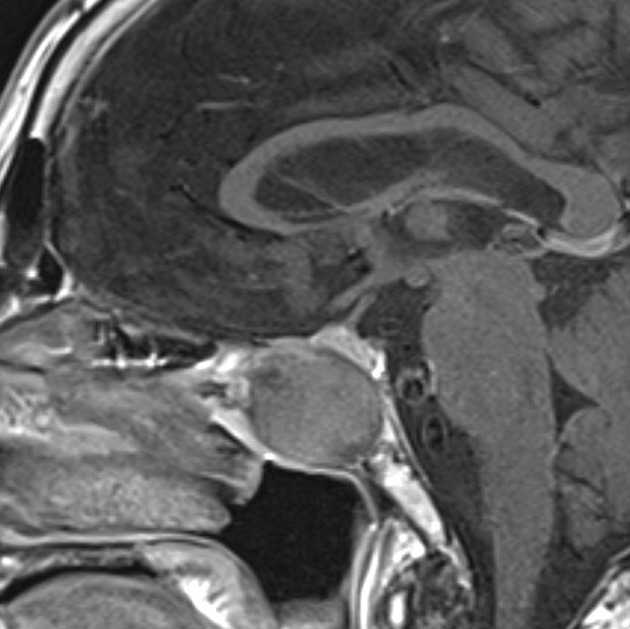
pituitary macroadenoma: ≥10 mm, sellar and suprasellar extension sometimes with snowman sign, remodeling of the sella
pituitary microadenoma: <10 mm, confined to the pituitary gland/sella, delayed/lower enhancement compared to the remaining gland
metastatic PitNET (pituitary carcinoma): rare, spread elsewhere in the CNS or body
pituicytoma: arise from the neurohypophysis and infundibulum, indolent, often absent pituitary bright spot, T2 hypo-isointense with prominent flow voids
granular cell tumor: arise from the posterior pituitary and infundibulum, can be indistinguishable from pituicytoma but often heterogeneously enhancing, hyperdense on CT
spindle cell oncocytoma: indistinguishable from macroadenoma, shows intense and early heterogenous enhancement
pituitary metastases: from breast, lung, kidney, gastrointestinal tract and nasopharynx 3 : involve the pituitary gland (grows rapidly: normal size pituitary fossa, osseous destruction) or the infundibulum (can have an absent bright spot)
pituitary lymphoma: rare, vivid possibly heterogenous enhancement, restricts diffusion
pituitary blastoma: rare, infants and young children
-
-
suprasellar/parasellar
meningioma: homogeneously enhancing, dural tail, hyperostosis, can narrow cavernous ICA
craniopharyngioma (adamantinomatous or papillary): heterogeneously enhancing sellar/suprasellar mass with cystic areas and calcifications, separate from a normal pituitary gland
hypothalamic/optic chiasmatic astrocytoma/glioma: T2 hyperintense, variable enhancement, often associated with neurofibromatosis type 1
hamartoma of tuber cinereum: follows grey matter on all sequences, gelastic seizures, precocious puberty 3
germinoma: hypercellular tumor (low ADC, hyperdense on CT), commonly in younger patients <20 years of age
dermoid/epidermoid/teratoma: restrict diffusion (epidermoid), incomplete FLAIR suppression (epidermoid)
-
sphenoid/clival
chordoma: T2 hyperintense, commonly in patients from 20-40, arise from the clivus
-
-
cellular infiltrates
-
Langerhans cell histiocytosis (most common)
-
hypophysitis including infundibuloneurohypophysitis
-
-
other lesions
berry aneurysm: parasellar/suprasellar, flow void, pulsation artifact, may be peripherally calcified
Rathke cleft cyst: intrasellar, non-enhancing cystic lesion, cyst with a dot sign
lipoma: suprasellar, fat signal lesion
mucocoele: centered in the sphenoid sinus, no solid enhancement
pituitary abscess: peripheral enhancing cystic lesion that restricts diffusion
pituitary stone: low signal, enlarged sella turcica
See also
It is also helpful to consider differentials narrowed by pattern of MRI appearance.


 Unable to process the form. Check for errors and try again.