
Tectal gliomas fall under the grouping of childhood brainstem gliomas and unlike the other tumors in that group they are typically low grade astrocytomas with good prognosis.
On this page:
Epidemiology
Tectal plate gliomas are encountered in children and adolescents 4. A male predilection has sometimes been reported although this is by no means certain 3.
Associations
An association with neurofibromatosis type I (NF1) has been reported 3,4.
Clinical presentation
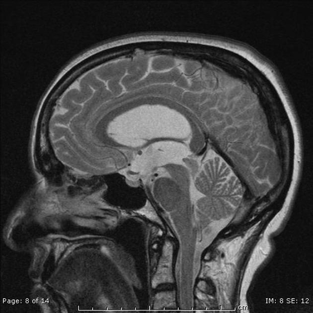
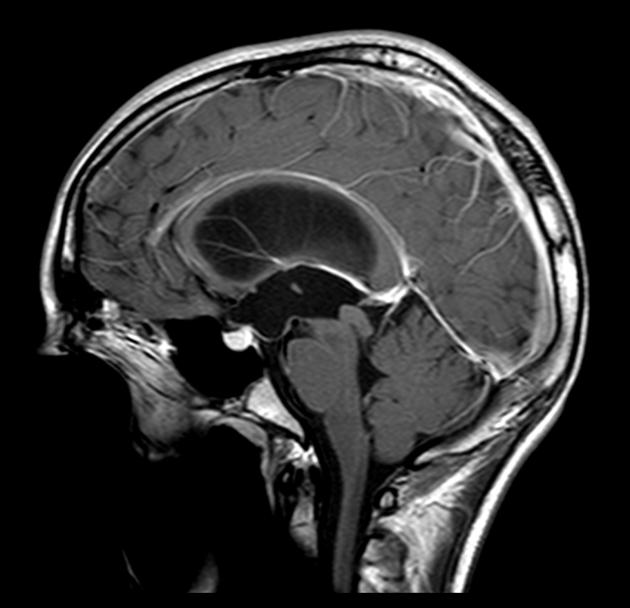
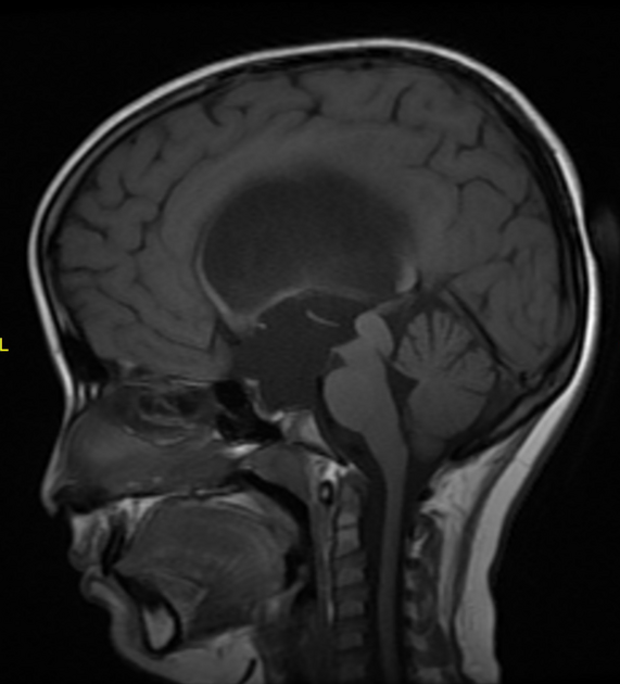
Presentation is commonly with headache as mass expansion of the tectum causes narrowing at the cerebral aqueduct and associated obstructive hydrocephalus 3,4.
Additional symptoms, secondary to compression of the medial longitudinal fasciculus, may include upward gaze palsy and diplopia (Parinaud syndrome), although this is less common 3,4.
Pathology
The vast majority of lesions are low grade astrocytoma, although occasionally other glial series tumors are encountered in the tectal region including ependymoma, ganglioglioma and primitive neuroectodermal tumors (PNET) 3.
Radiographic features
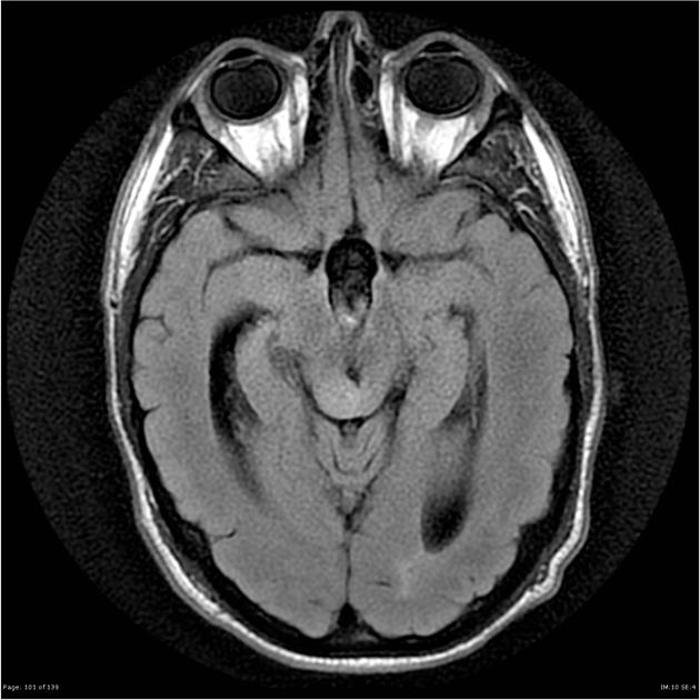
CT
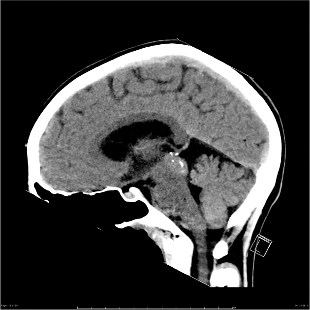
Typical CT finding is homogeneous expansion of tectal plate, isodense to grey matter with minimal enhancement on postcontrast images 1,3. On CT it is not uncommon to find a central tectal calcification 2,3.
MRI
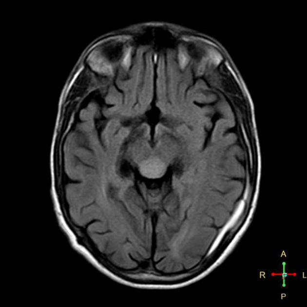
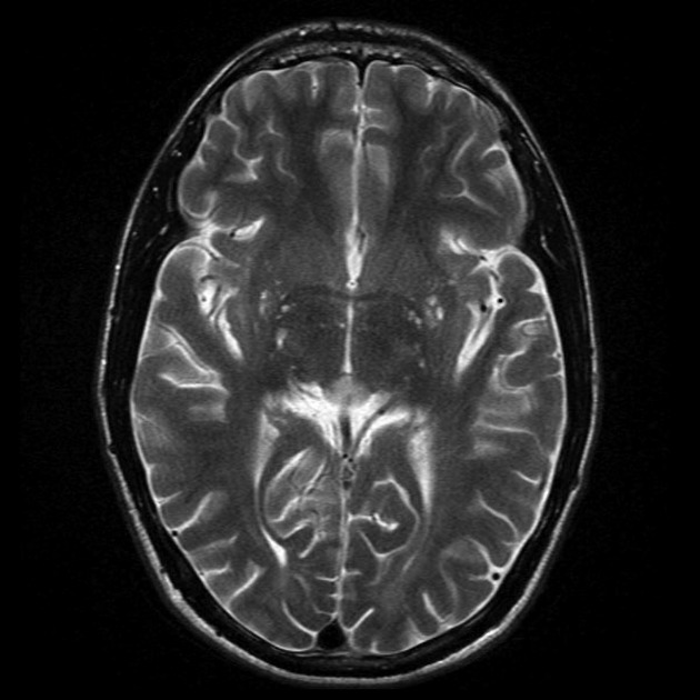
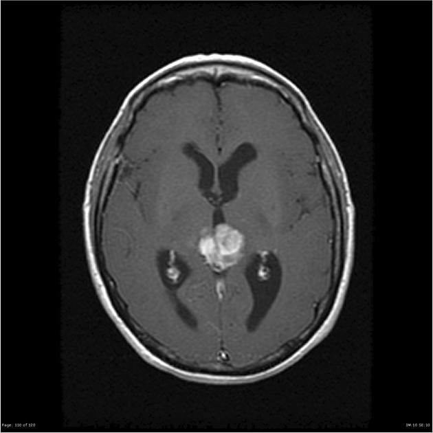
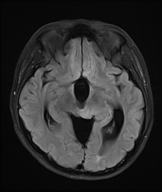
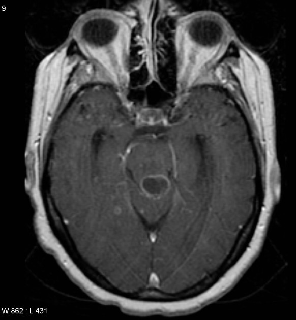
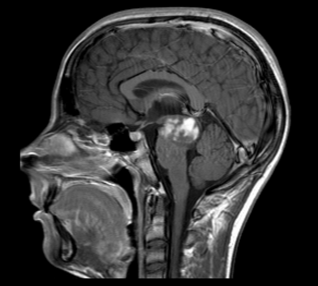
Typically the tumors demonstrate expansion of the tectal plate by a solid nodule of tissue.
T1: iso to slightly hypointense to grey matter 1-3
T2: hyperintense to grey matter
T1 C+ (Gd): usually no enhancement
With time the mass can develop small cystic spaces (sometimes associated with neurological deficits) or calcification 3.
Higher grade tumors tend to be larger and tend to enhance more vividly 3.
Treatment and prognosis
As tectal plate gliomas are low grade and often very slow growing, shunting is often the only required intervention for long term survival. As surgical biopsy can have significant morbidity in this area, usually the diagnosis is made on imaging findings alone.
In the minority of patients who progress, radiotherapy often leads to local control or even tumor regression 2. Surgical excision is sometimes necessary 3.
Imaging predictors of patients who will need further treatment include a size greater than 2.5 cm and presence of contrast enhancement 3.
Differential diagnosis
When the tectum is near-normal then the differential is largely limited to:
-
no mass lesion
a focal stenosis or web may be visible
With larger lesions, where the mass is not definitely arising from the tectal plate then the differential is essentially that of a pineal region mass and therefore includes:
In patients with NF1 a hamartoma should also be considered. They tend to have some T1 hyperintensity 4.


 Unable to process the form. Check for errors and try again.