
Cruciate ligament of the atlas
Citation, DOI, disclosures and article data
At the time the article was created Aaron Wong had no recorded disclosures.
View Aaron Wong's current disclosuresAt the time the article was last revised Arlene Campos had no financial relationships to ineligible companies to disclose.
View Arlene Campos's current disclosures- Cruciform ligament (atlas)
- Cruciate ligament of atlas
- Transverse atlantal ligament
- Transverse atlantic ligament
- Cruciform ligaments (atlas)
- Transverse atlantic ligaments
- Transverse atlantal ligaments
- Cruciate ligaments of atlas
- Transverse band (cruciate ligament)
- Longitudinal band (cruciate ligament)
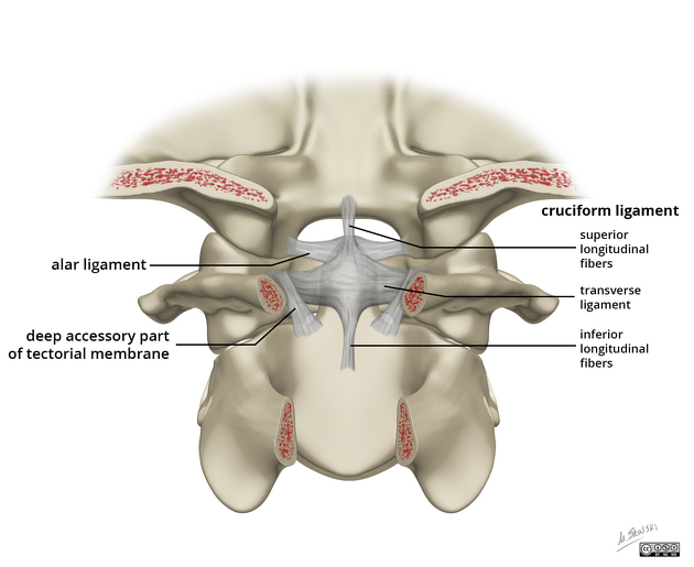
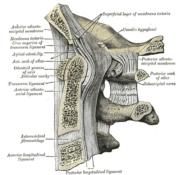
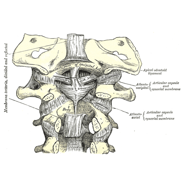
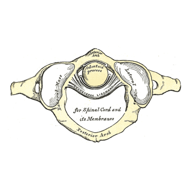
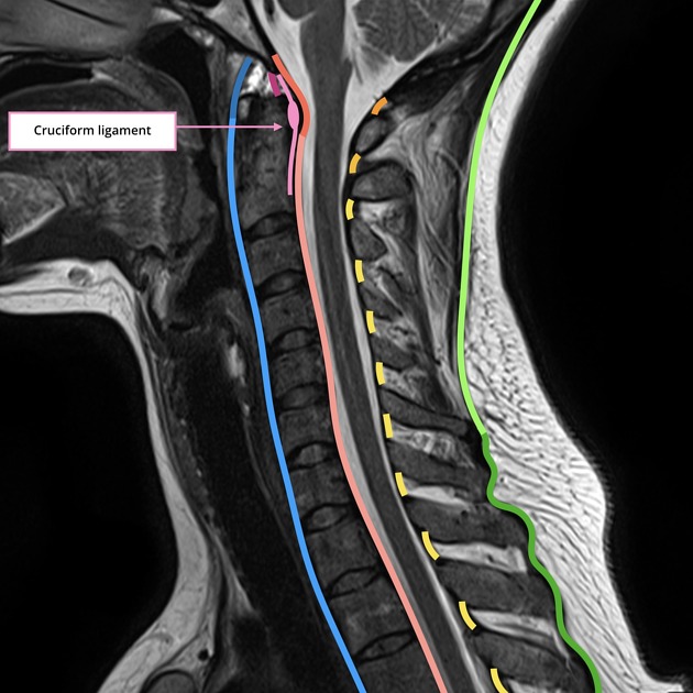
The cruciate ligament of the atlas (also known as the cruciform ligament) is an important ligamentous complex that holds the posterior dens of C2 in articulation at the median atlantoaxial joint. It lies behind a large synovial bursa (surrounded by loose fibrous capsule) and consists of two bands:
-
longitudinal band
attaches the body of the C2 (axis) to the clivus and foramen magnum in the midline, lying between the apical ligament and tectorial membrane
relatively weak and hence does not contribute any significant stability
-
transverse band (also known as the transverse atlantic or atlantal ligament)
attaches to a small tubercle on the medial cortex of the C1 (atlas) lateral masses on both sides anterior to the tectorial membrane and dura
passes posterior to the dens, with a small intervening synovial capsule, fixing the dens to the posterior margin of the anterior arch of the atlas
strongest spinal ligament 2
With the alar ligament, the transverse band is the primary stabilizer of the atlantoaxial joint 2.
References
- 1. McMINN. Lasts Anatomy Regional and Applied. CHURCHILL LIVINGSTONE. (2003) ISBN:B0084AQDG8. Read it at Google Books - Find it at Amazon
- 2. Offiah CE, Day E. The craniocervical junction: embryology, anatomy, biomechanics and imaging in blunt trauma. Insights into imaging. 8 (1): 29-47. doi:10.1007/s13244-016-0530-5 - Pubmed
Incoming Links
- Retro-odontoid pseudotumour
- Atlas (C1)
- Apical ligament
- Anderson and D'Alonzo classification of odontoid process fracture
- Whiplash syndrome
- Gehweiler classification of atlas fractures
- Atlanto-occipital articulation
- Atlanto-axial articulation
- Alar ligament
- AO Spine classification of upper cervical injuries
- Jefferson fracture
- Tectorial membrane of the spine
- Os odontoideum
- Supraodontoid space
- Rule of Spence
- Crowned dens syndrome
- Cervical spine ligaments
- Axis (C2)
Related articles: Anatomy: Spine
-
osteology[+][+]
- vertebrae
- spinal canal
- cervical spine
- thoracic spine
- lumbar spine
- sacrum
- coccyx
-
anatomical variants
- vertebral body
- neural arch
- transitional vertebrae
- ossicles
- ossification centers
- intervertebral disc[+][+]
- articulations[+][+]
- ligaments
- musculature of the vertebral column[+][+]
- muscles of the neck
- muscles of the back
-
suboccipital muscle group
- rectus capitis posterior major muscle
- rectus capitis posterior minor muscle
- obliquus capitis superior muscle
- obliquus capitis inferior muscle
- splenius capitis muscle
- splenius cervicis muscle
- erector spinae group
- transversospinalis group
- quadratus lumborum muscle
-
suboccipital muscle group
- spinal meninges and spaces[+][+]
-
spinal cord[+][+]
- gross anatomy
-
white matter tracts (white matter)
- corticospinal tract
- anterolateral columns
- lateral columns
-
dorsal columns
- fasiculus gracilis (column of Goll)
- fasiculus cuneatus (column of Burdach)
- grey matter
- nerve root
- central canal
- functional anatomy
- spinal cord blood supply
- sympathetic chain[+][+]


 Unable to process the form. Check for errors and try again.