
Umbilical arterial (UA) Doppler assessment is used to survey fetal well-being in the third trimester of pregnancy. Abnormal umbilical artery Doppler is a marker of placental insufficiency and consequent intrauterine growth restriction (IUGR) or suspected pre-eclampsia.
Umbilical artery Doppler assessment has been shown to reduce perinatal mortality and morbidity in high-risk obstetric situations 5.
As a general rule, a degree of caution should be exercised with the routine use of Doppler in pregnancy, due to the concerns related to heating/thermal effects from the high intensities of Doppler ultrasound.
On this page:
Indications
Umbilical Doppler assessment is indicated in scenarios where there is a risk of fetal growth restriction or poor perinatal outcome. It is also used to stage twin-twin transfusion 7.
Doppler ultrasound evaluation of the fetoplacental circulation is not indicated in low-risk pregnancies 7.
Maternal conditions
Pregnancy-related conditions
suspected IUGR
previous pregnancy with IUGR or fetal death in utero
Radiographic features
Ultrasound
The spectral Doppler indices measured at the fetal end, the free loop, and the placental end of the umbilical cord are different with the impedance highest at the fetal end. The changes in the indices are likely to be seen at the fetal end first. Ideally, the measurements should be made in the free cord, however, for consistency of recording in cases being followed up, a fixed site would be more appropriate, i.e. fetal end, placental end, or intra-abdominal portion. Due to difficulty with measuring the cord at the fetal end in many growth-restricted fetuses, measurement in a free loop is acceptable 7.
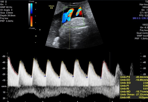
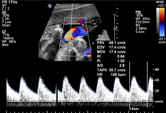
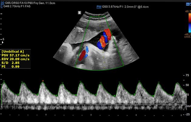
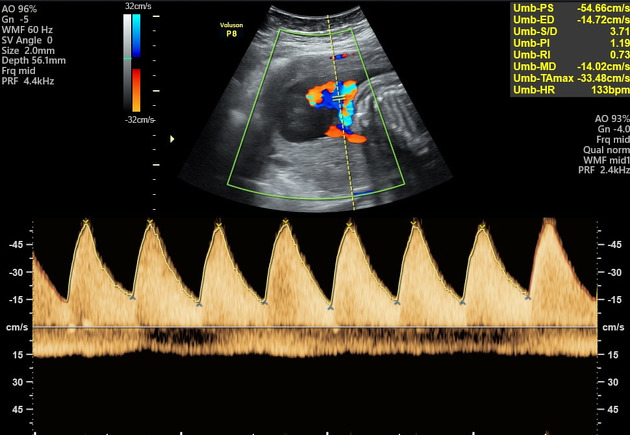
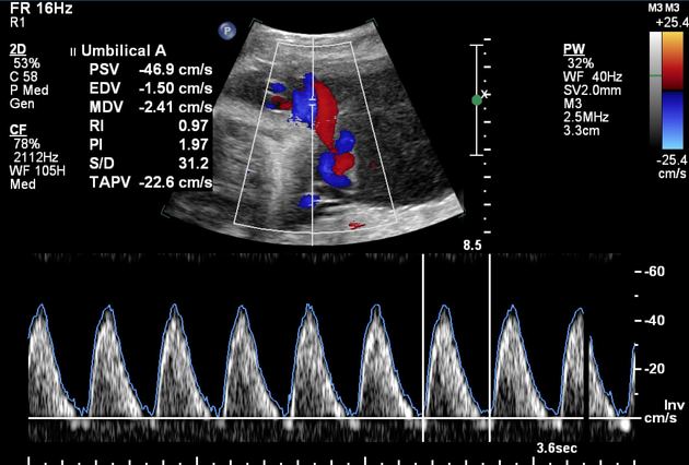
Waveform
The umbilical arterial waveform usually has a "sawtooth" pattern with flow always in the forward direction, that is towards the placenta. An abnormal waveform shows absent or reversed diastolic flow. Before the 15th week, the absence of diastolic flow may be a normal finding 6.
The 95% confidence interval limit slowly decreases for both the resistive index (RI) and pulsatility index (PI) through the course of gestation due to progressive maturation of the placenta and increase in the number of tertiary stem villi.
Parameters
The commonly used parameters are:
umbilical arterial S/D ratio (SDR): systolic velocity / diastolic velocity
pulsatility index (PI) (Gosling index): (PSV - EDV) / TAV
resistive index (RI) (Pourcelot index): (PSV - EDV) / PSV
TAV: time-averaged velocity
The Doppler indices have been found to decline gradually with gestational age (i.e. there is more diastolic flow as the fetus matures):
-
S/D ratio mean value decreases with fetal age 8
at 20 weeks, the 50th percentile for the S/D ratio is 4
at 30 weeks, the 50th percentile is 2.83
at 40 weeks, the 50th percentile is 2.18
RI mean value decreases from 0.756 to 0.609
PI mean value decreases from 1.270 to 0.967
Classification
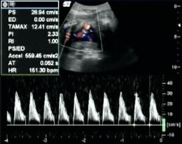
In growth-restricted fetuses and fetuses developing intrauterine distress, the umbilical artery blood velocity waveform usually changes in a progressive manner as below
reduction in end-diastolic flow: increasing RI values, PI values, and S/D ratio
absent end-diastolic flow (AEDF): RI = 1
Further assessment tools
Abnormal umbilical artery Doppler is an indication of further sonographic workup of the degree of placental insufficiency:
See also
External links
If any of these links are broken or for other problems and questions, please contact editors@radiopaedia.org.


 Unable to process the form. Check for errors and try again.