
The ankle horizontal beam lateral view is a modified lateral view part of a three view ankle series.
On this page:
Indications
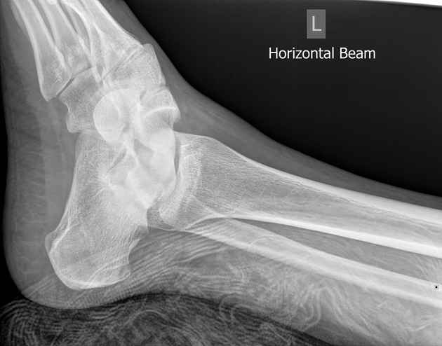
This projection is used to assess the distal tibia and fibula, talus, navicular, cuboid, the base of the 5th metatarsal and calcaneus. It is a highly adaptable projection that can be used in trauma or with patients who are unable to ambulate to the desired standard lateral position.
Patient position
patient is in a supine position
-
this projection can be done from either side depending on the makeup of the room and the patients' pathology
if projection is mediolateral, the non-affected leg is raised/placed on a stand in a flexed position to avoid superposition
if projection is lateromedial, both legs can lay in their natural AP position
foot in dorsiflexion if possible
Technical factors
mediolateral/lateromedial horizontal beam projection
-
centering point
-
mediolateral
bony prominence of the medial malleolus of the distal tibia
-
lateromedial
bony prominence of the lateral malleolus of the distal fibula
-
-
collimation
-
orientation
portrait or landscape
-
detector size
18 cm x 24 cm
-
exposure
50-60 kVp
3-5 mAs
-
SID
100 cm
-
grid
no
Image technical evaluation
The distal fibula should be superimposed by the posterior portion of the distal tibia.
The talar domes should be superimposed allowing for adequate inspection of the superior articular surface of the talus.
The joint space between the distal tibia and the talus is open and uniform.
Practical points
In situations where the patient cannot be moved, this projection can be invaluable as it requires little to no patient movement and can be replicated in ICU wards.
If possible, placing a radiolucent immobilization sponge under the ankle in question will prevent any artifacts from bed/pillows that are native to trauma rooms.
Superior-inferior malalignment of the superior aspect of the talus is resultant of the tibia not lying parallel to the image receptor. To adjust this, angle the tube superior-inferior to mimic the tibia laying parallel. This is not ideal but in trauma, it may be the only option.
Anterior-posterior malalignment of the talar domes is due to over or under rotation of the foot. To adjust this, angle the tube anterior-posterior to mimic correct positioning. This is not ideal but in trauma, it may be the only option.


 Unable to process the form. Check for errors and try again.