
The AP oblique rib projection is performed to best demonstrate the axillary ribs. Oblique ribs may be conducted either as an anterior oblique or posterior oblique view.
On this page:
Indications
The AP oblique view specifically focuses on the axillary ribs. The rib series is often considered to be an unnecessary, unjustified projection in many radiology departments. Indeed the Royal College of Radiologists (UK) iRefer guidelines state "Demonstration of a simple rib fracture does not usually alter management but if a complication such as pneumothorax or infection is suspected, chest radiograph would be appropriate" 3. Thus if the projection might change the patient management it may still be considered pertinent and worthy of discussion.
Other indications for chest oblique views are assessing the pathology in the lung fields, trachea, mediastinal structures, contours of heart and large vessels 2.
Patient position
the patient may be erect or supine with their right (RPO) or left posterior (LPO) side closest to the image receptor 2
affected side is rotated 45 degrees towards the IR 2
the patient’s arm closest to the receptor is raised and placed on their head, with the other on their hip 2
Technical factors
anteroposterior oblique projection
-
respiration
suspended inspiration
-
centering point
above diaphragm: level of T7 (8 to 10 cm below the vertebral prominens. )
midway between the midsagittal plane and lateral margin of thorax 2
-
collimation
medially include 5 cm lateral to the sternoclavicular joint of the unaffected side
laterally to the lateral rib margin
superoinferiorly above diaphragm 5 cm above sterno-clavicular joint
superoinferiorly below diaphragm lower costal margin
-
orientation
portrait
-
detector size
35 cm x 43 cm 2
-
exposure
110 – 125 kVp 2
12 – 20 mAs
-
SID
183 cm 2
-
grid
yes
Image technical evaluation
-
above diaphragm
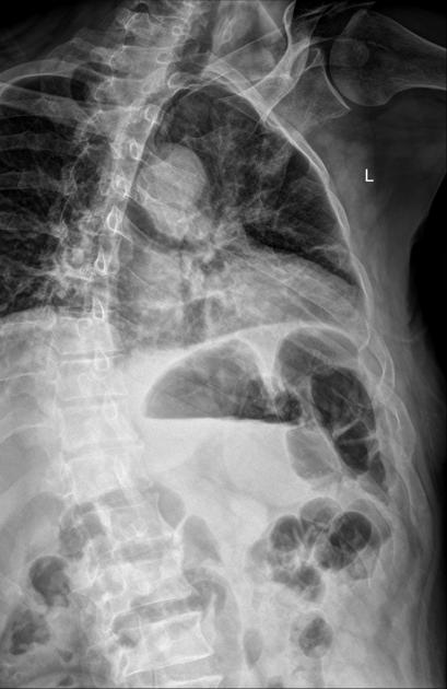
1st – 10th axillary ribs of the affected side are demonstrated without superimposition
thoracic vertebrae are included
Practical points
If adequate obliquity has been achieved, the glenohumeral joint on the affected side will be open.
If you are confused by which way to oblique the patient depending on AP/PA position, remember that the spinous processes will always point towards the unaffected side (i.e. for left oblique ribs, spinous processes point to the right) (see Case 1).
The antomy shown in LPO position is equivalent to RAO position while the anatomy shown in RPO position is equivalent to LAO position 2.
Conducting AP rib oblique views produces less magnification of the ribs, and provides more bony detail than that of the PA view 1.
When a patient is unable to stand for an erect projection, have the patient supine and supported by immobilization devices to adjust the patient into an oblique position.
For the PA oblique projection (LAO/RAO), the affected side is rotated 45 degrees away from the IR, with the CR as per the AP oblique view.


 Unable to process the form. Check for errors and try again.