
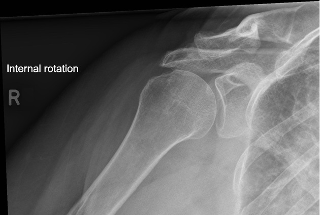
The shoulder internal rotation view is an additional projection to the standard shoulder series it is often combined with the external rotation view to visualize the entirety of the humeral head.
On this page:
Indications
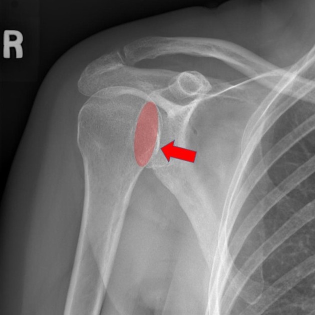
This projection shows the lesser tubercle of the humerus in profile and can be used to detected suspected Hill-Sachs lesions.
Patient position
- patient is preferably erect
- the midcoronal plane of the patient is parallel to the image receptor, in other words, the patient's back is against the image receptor
- the glenohumeral joint of the affected side is at the center of the image receptor
- the affected arm is internally rotated
- the patient is slightly rotated 5-10°. Therefore, the body of the scapula is laying parallel with the image receptor
- some departments may require further rotation to mimic that of 'true AP' projection
Technical factors
- anteroposterior projection
-
centering point
- 2.5 cm inferior to the coracoid process, or 2 cm inferior to the lateral clavicle at the level of the glenohumeral joint
-
collimation
- superior to the skin margins
- inferior to include one-third of the proximal humerus
- lateral to include the skin margin
- medial to include 2/3 of the clavicle
-
orientation
- landscape
-
detector size
- 24 cm x 30 cm
-
exposure
- 60-70 kVp
- 10-18 mAs
-
SID
- 100 cm
-
grid
- yes (this can vary departmentally)
Image technical evaluation
- 2/3 of the clavicle is visible
- overlap of the humeral head with the glenoid
- no foreshortening of the scapular body (as per the patient rotation discussed in the positioning)
- the lesser tubercle of the humerus is in profile
Practical points
The technical factors of this examination are not particularly demanding, and there is not much room for positioning error other than over or under rotation to compensate for the scapular body.
Patients find it hard to rotate their arm internally, so take this into consideration and only have them perform is at the last minute.
An open glenohumeral joint is a sign of over-rotation toward the affected side. This results in a more ‘glenoid AP’ view; some departments may ask the projection be done in the 'glenoid AP' view.


 Unable to process the form. Check for errors and try again.