
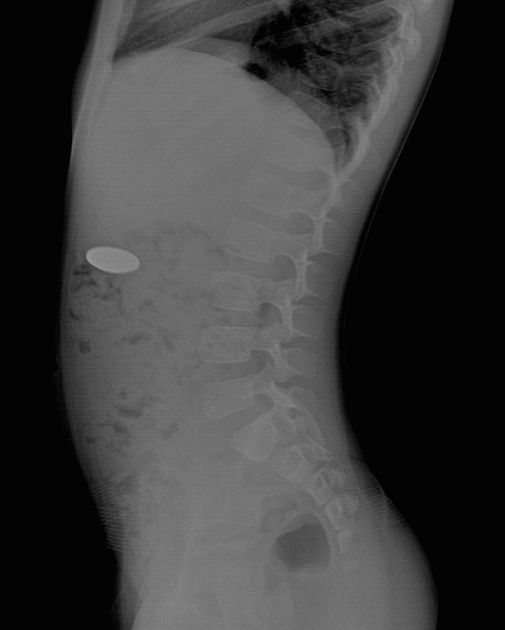
Abdominal radiography can be useful in many settings. Before the advent of CT, it was a primary means of investigating gastrointestinal pathology and often allowed indirect evaluation of other abdominal viscera.
On this page:
Indications
Although abdominal radiography has lower sensitivity and specificity than a CT of the abdomen, it still serves a role as an adjunct or optional test. Uses for abdominal radiography include:
-
a preliminary evaluation of bowel gas in an emergent setting
a negative study in a low pretest probability patient may obviate the need for a CT study and therefore lower radiation dose
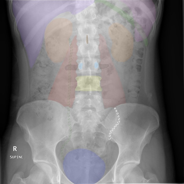
evaluation of radiopaque tubes and lines
evaluation for radiopaque foreign bodies
evaluation for postprocedural intraperitoneal/retroperitoneal free gas
monitoring the amount of bowel gas in postoperative ileus
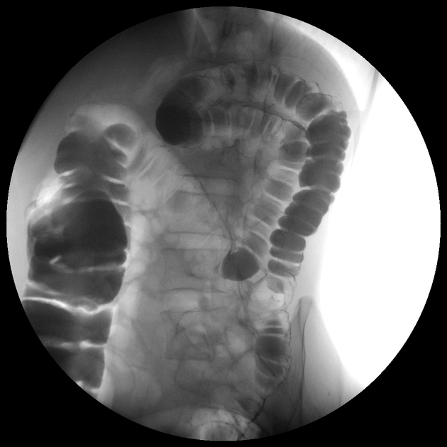
monitoring the passage of contrast through the bowel
monitoring renal calculi
Contraindications
-
pregnancy is a relative contraindication to the use of ionizing radiation
non-ionizing studies (e.g. ultrasound or MRI) should be tried first
abdominal radiographs administer a much lower radiation dose than CT
Projections
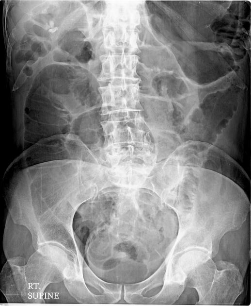
Standard projections
-
can be performed as a standalone projection or as part of an acute abdominal series
-
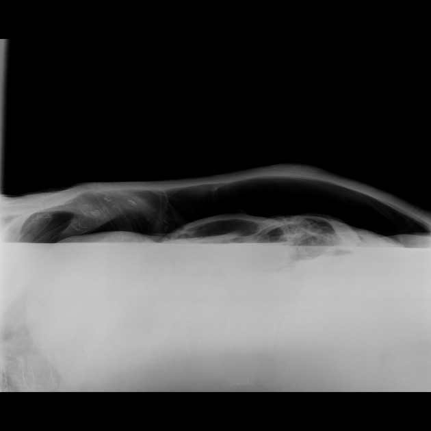
often taken with the supine view, when used together it is a valuable projection in assessing gas-fluid levels, and free gas in the abdominal cavity.
Additional projections
Generally, plain radiograph examination of the abdomen comprises an AP supine and PA erect view, supplemented by a number of additional views as clinically indicated.
NB: please note that in the UK, a single supine abdominal radiograph is the norm, i.e. erect abdominal x-rays have not been routinely performed for decades 2.
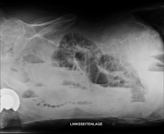
-
performed as an alternative to the PA erect view to assess for free gas in the abdominal cavity
-
often used as a problem solving view during the identification and localization of foreign bodies
-
performed if the patient is unable to lie supine
-
used when it is unsafe to perform both a PA erect or a lateral decubitus view, this projection requires no patient movement.
-
used in barium studies and the location of foreign bodies and/or lines such as a Tenckhoff catheter
Procedure
Preprocedural evaluation
The patient should be gowned with minimum clothing. Radiopaque materials (zippers, belts, etc.) should be removed.
If relevant, enteric tube suction should be avoided before the study. Ideally, the patient's bladder should be emptied as well.
Technique
Abdominal radiographs may be obtained in the radiology department or may be performed portably. Views should generally include either the diaphragm or inferior pubic ramus. Gonadal shielding may be provided for men.
Portable abdominal radiographs may be necessary due to patient immobility but are of much poorer quality.
kVp
The kVp of the x-ray beam may be altered in order to bring out different aspects of the abdominal radiograph:
lower kVp offers greater tissue contrast and better visualization of gas, but there is decreased penetration of the x-ray beam
higher kVp may be useful for evaluation of radiopaque objects (contrast, tubes, lines, etc.)


 Unable to process the form. Check for errors and try again.