
The carpal bridge view an additional view of the three view series of the wrist and carpal bones. It is a specialized projection that involves keeping the patient's wrist in flexion.
On this page:
Indications
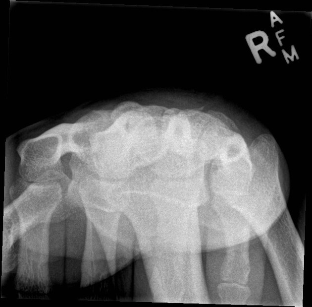
The carpal bridge view is requested to assess the dorsal aspect of the scaphoid, lunate and the triquetrum.
Patient position
- the patient is seated alongside the table
- dorsal aspect of affected wrist is placed on the detector in flexion
- flex the wrist as much as tolerable to the patient
Technical factors
- tangential projection
-
centering point
- midcarpal region
- the central ray is angled approximately 45 degrees to the long axis of the forearm
-
collimation
- laterally to the skin margins
- dorsal to the skin margins
- ventral to the carpometacarpal joint
-
orientation
- portrait
-
detector size
- 18 cm x 24 cm
-
exposure
- 50-60 kVp
- 3-5 mAs
-
SID
- 100 cm
-
grid
- no
Image technical evaluation
There should be a clear outline of the dorsal aspect of the carpal bones with no superimposition.
Practical points
This is a very specialized projection and it can cause the patient significant pain if not performed properly, yet it can be very helpful in imaging triquetrum fractures. It is best to demonstrate to the patient physically what you plan to do before making them perform it, this way they are not in discomfort for long.


 Unable to process the form. Check for errors and try again.