
The clavicle AP cephalic angulation view is a standard projection part of the clavicle series and is often used in conjunction with the AP clavicle view.
On this page:
Indication
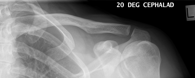
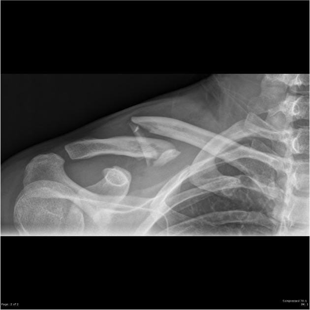
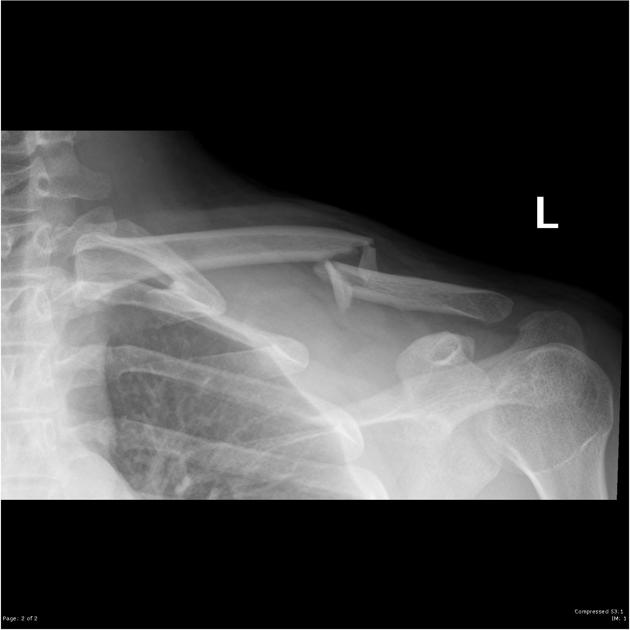
This projection straightens out the clavicle and projects most of it above the scapula and second and third rib. It can help to determine angulation of fractures and dislocation 1.
Patient position
- patient is preferably erect
- midcoronal plane of the patient is parallel to the image receptor, in other words, the patient's back is against the image receptor
- the clavicle of the affected side is at the center of the image receptor
- affected arm is in a neutral position
Technical factors
- anteroposterior projection
-
centering point
- just below mid clavicle
- angled cephalic 15-30°
-
collimation
- superior to the skin margins
- inferior to include mid scapula
- lateral to include the skin margin
- medial to include the sternoclavicular joint
-
orientation
- landscape
-
detector size
- 18 cm x 24 cm
-
exposure
- 60-70 kVp
- 10-18 mAs
-
SID
- 100 cm
-
grid
- yes (this can vary departmentally)
Image technical evaluation
- the clavicle is 'flattened out' projecting above the shoulder girdle
- a slight overlap of the humeral head within the glenoid
Practical points
Remember to move your detector to compensate for the cephalic angulation. This projection can often better demonstrate subtle clavicle fractures, if you can't see anything on the AP projection, this should be your next point of call.


 Unable to process the form. Check for errors and try again.