
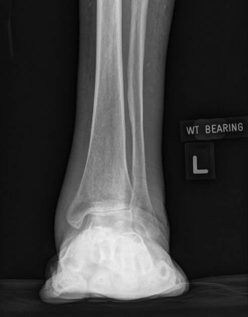
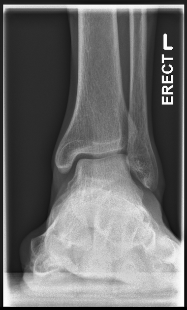
The weight-bearing AP view of the ankle is a specialized projection that places the joint under normal weight-bearing conditions. The projection is utilized to assess the joint under stress and better demonstrate structural and functional deformities.
On this page:
Indications
This projection is utilized to assess the structural integrity of the ankle joint. If the patient is able, weight-bearing views should be performed in acute and follow up settings 1.
Other indications include:
- assessment of fragment position and implants in postoperative follow up
- evaluation of fracture healing
- evaluation of tibiofibular clear space and overlap in suspected syndesmotic injury
- evaluation of hindfoot deformities
In addition, this view can show bony diseases or lesions of the distal lower leg, talus and proximal fifth metatarsal.
Ultimately the radiographer will determine if the projection is safe to perform.
Patient position
- the patient is standing on an upright stand with the ankle in question central and perpendicular to the detector (toes facing the x-ray tube)
- there is no rotation, the foot is in a neutral rotation
- the patient is distributing weight evenly
- have something for the patient to hold onto, especially if they unsteady on their feet
Technical factors
- anteroposterior projection
-
centering point
- the midpoint of the lateral and medial malleoli
-
collimation
- laterally to the skin margins
- superior to examine the distal third of the tibia and fibula
- inferior to the proximal aspect of the metatarsals
-
orientation
- portrait
-
detector size
- 24 cm x 30 cm
-
exposure
- 50-60 kVp
- 3-5 mAs
-
SID
- 100 cm
-
grid
- no
-
grid
- no
Image technical evaluation
- the distal fibula should be slightly superimposed the distal tibia (>10mm)
- the lateral and medial malleoli of the distal fibula and tibia are in profile
- the tibiotalar joint space should be open, yet the full mortise joint should not be visualized on the AP
Useful measurements
Practical points
Although this projection is helpful to assess lower limb injuries better, it must be performed safely. Explain and demonstrate to the patient how it is performed. Give them something to hold on to. If the exam does not feel safe, explore alternative methods such as a standard, supine projection.


 Unable to process the form. Check for errors and try again.