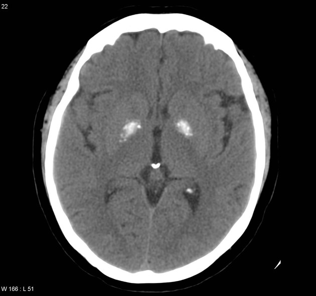
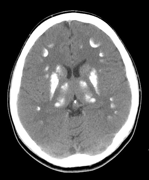
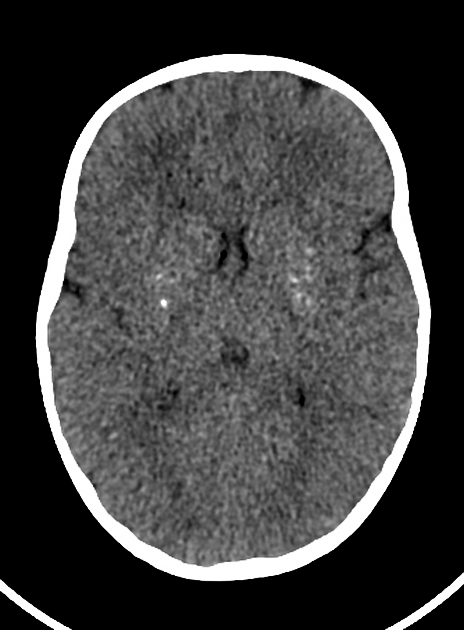
Basal ganglia calcification is common and is seen in approximately 1% of all CT scans of the brain, depending on the demographics of the scanned population. It is seen more frequently in older patients and is considered a normal incidental and idiopathic finding in an elderly patient but should be considered pathological in persons younger than the age of 40 years unless proved otherwise 5.
6. Livingston JH, Stivaros S, Warren D, Crow YJ. Intracranial calcification in childhood: a review of aetiologies and recognizable phenotypes. (2014) Developmental medicine and child neurology. 56 (7): 612-26. doi:10.1111/dmcn.12359 - Pubmed
7. S. Dehkharghani, W.P. Dillon, S.O. Bryant, N.J. Fischbein. Unilateral Calcification of the Caudate and Putamen: Association with Underlying Developmental Venous Anomaly. (2010) American Journal of Neuroradiology. 31 (10): 1848. doi:10.3174/ajnr.A2199 - Pubmed
8. Verulashvili IV, Glonti LSh, Miminoshvili DK,et al. [Basal ganglia calcification: clinical manifestations and diagnostic evaluation]. (2006) Georgian medical news. Pubmed


 Unable to process the form. Check for errors and try again.