
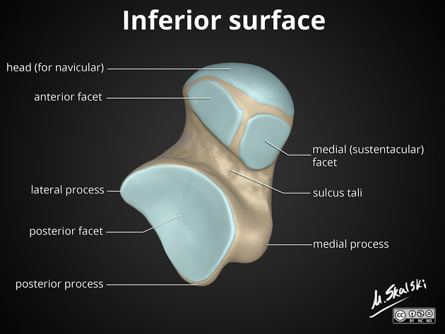
The Broden's view (or Broden view) is a specialized projection that accurately 1 examines the large posterior calcaneal facet and the subtalar joint 2.
As technology advances, computed tomography (CT) has widely been used to better visualize and characterize fragment displacements and fracture lines. Yet, there remain various reasons for plain film imaging being the choice modality, like inaccessibility or absence of a CT scanner in rural regions and the affordability of the scans for patients.
Depending on departmental protocol, one or four images at varying cephalic angles may be acquired to view different parts of the posterior calcaneal facet 3. The cephalic angles 4 recommended are at:
10°: demonstrates most posterior aspect
20°: default angle used if only one single image is allowed
30°
40°: demonstrates most anterior aspect
On this page:
Indications
This view at varying angles aid in detecting potential fracture displacement, depression or subluxation of the hindfoot after a notable 2:
axial loading mechanism (for intra-articular fractures)
low energy mechanism (for extra-articular fractures)
Patient position
patient is supine or seated with the affected limb extended
the posterior aspect of the ankle is resting on the image receptor
-
the affected leg must be rotated 45° internally
internal rotation must be from the hip; isolated inversion of the ankle will result in a non-diagnostic image
foot should be in neutral dorsiflexion
Technical factors
anteroposterior axial projection
-
centering point
the beam is angled at 10°, 20°, 30° and 40° cephalad from the long axis of the foot
central ray is placed over the lateral malleolus 2
-
collimation
lateral to the skin margins
superior to the distal third of tibia and fibula
inferior to the tarsals
-
orientation
portrait
-
detector size
18 cm x 24 cm
-
exposure
55-60 kVp
3-5 mAs
-
SID
100 cm
-
grid
no
Image technical evaluation
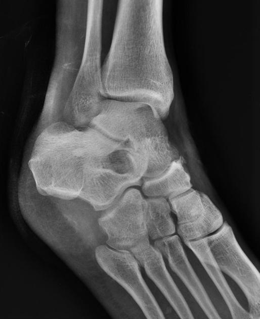
Clear visualization of the anterior and posterior talocalcaneal joint 5.
Practical points
Although having the patient's foot in dorsiflexion is preferred, many times this will not be possible due to pain. In such scenarios, it is equally effective to:
increase the cephalic angle to compensate for lack of dorsiflexion
raise the distal part of the leg (placing an immobilization sponge underneath the leg), ensuring the knee joint is kept extended


 Unable to process the form. Check for errors and try again.