
The cervical spine flexion and extension views demonstrate the seven vertebrae of the cervical spine when the patient is in a lateral position.
On this page:
Indications
These views are specialized projections often requested to assess for spinal stability.
Note, such functional views should not be performed on trauma patients without the strict instructions of a qualified clinician.
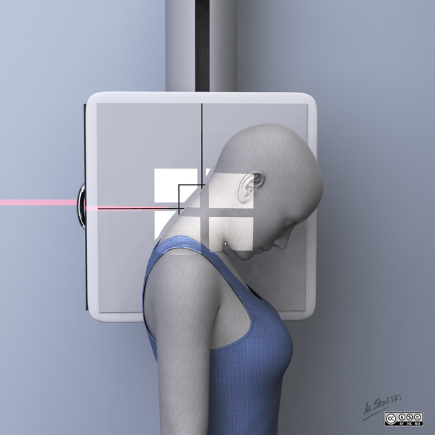
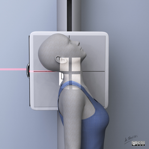
Patient position
the patient is erect, left side against the upright detector
the detector is placed portrait, parallel to the long axis of the cervical spine on the patients left side
the patient will have the neck in the extended (chin up) or flexion (chin down) position depending on the projection
Technical factors
lateral projection
-
centering point
2.5 cm above the jugular notch at the level of C4
-
collimation
superior to C1
inferior to T1
anterior to include soft tissue
posterior to the soft tissue
-
orientation
portrait
-
detector size
24 cm x 30 cm
-
exposure
50-75 kVp
20-50 mAs
-
SID
150-180 cm
-
grid
yes
Image technical evaluation
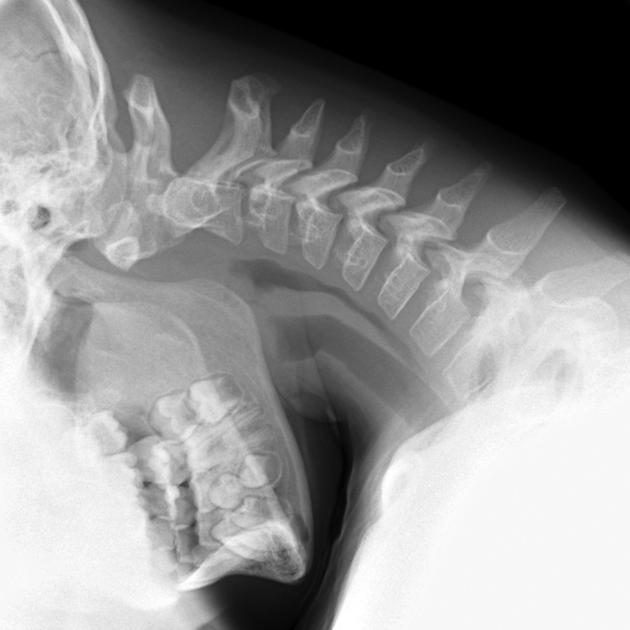
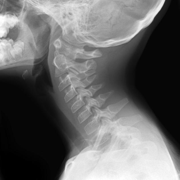
there should be clear visualization of C1 to T1
the image is labeled as 'flexion' or 'extension'
flexion images should demonstrate well separated spinous process
extension images should demonstrate crowding of the spinous process
Practical points
demonstrate to the patient what flexion and extension is before performing
ensure the patient is aware when the examination is over as to avoid extended periods of time in that position
patients who feel unstable on their feet can sit in a chair for this examination
ensure this radiographic series is safe to perform, i.e. part of a secondary survey or under the guidance of an authorized physician
>2.5-3.5 mm of intersegmental translation (a summation of the displacement observed between vertebra tracing the posterior line on both the flexion and extension view) is considered a marker of instability 3


 Unable to process the form. Check for errors and try again.