
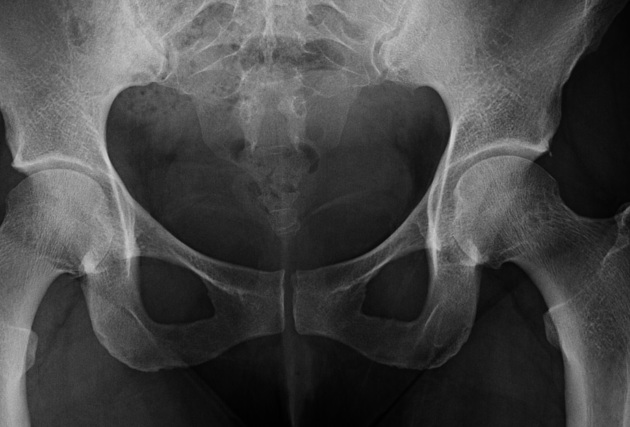
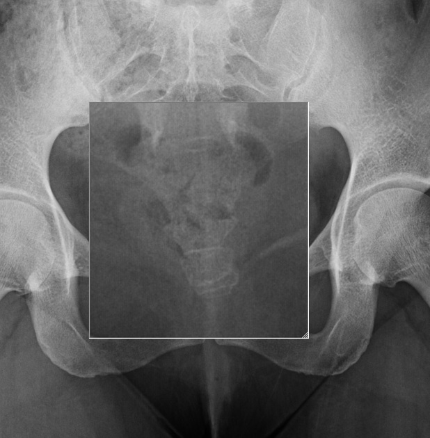
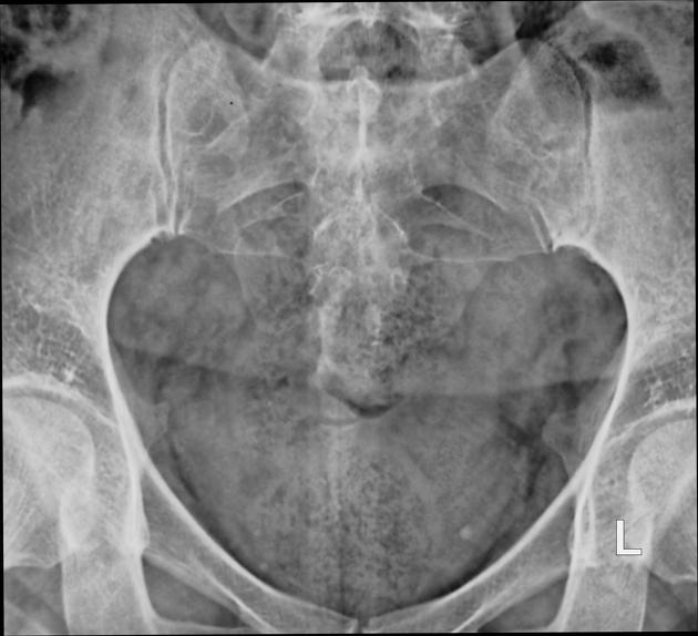
The coccyx anteroposterior (AP) view is used to demonstrate the coccyx, in conjunction with the sacrum and coccyx (lateral view). Follow departmental protocol in relation to imaging this region.
On this page:
Indications
This projection helps to visualize the pathology of the coccyx, especially fractures. To minimize superimposition of structures over the coccyx region, the urinary bladder and large colon should ideally be emptied before this examination 1.
Given that management of coccygeal fractures is nearly always non-operative, some radiology literature suggests that x-ray evaluation for coccygodynia is a waste of resources and exposes patients to unnecessary ionizing radiation, without having a measurable impact on clinical outcome. Thus, in some territories (e.g. the UK), the usual practice is to not perform routine imaging of the coccyx 2.
Patient position
the patient is supine, with arms placed comfortably by their side, legs extended 1
Technical factors
anteroposterior view
-
centering point
5 cm superior to the pubic symphysis at the mid-sagittal plane 1
-
central ray
angled 10° caudal 1
-
collimation
must adhere to the ALARA principle given the radiosensitive region exposed via the primary beam
close collimation to the area of interest
-
orientation
portrait
-
detector size
24 x 30 cm
-
exposure
80 kVp
15 mAs
-
SID
110 cm
-
grid
yes
Image technical evaluation
adequate penetration should clearly demonstrate the coccyx region
the coccyx is free of superimposition from the pubic rami
lateral margin of the coccyx is equidistant from the pelvic brim indicating no patient rotation
Practical points
given the proximity of this anatomy to the gonadal region, the risk versus benefit of ionizing radiation and diagnostic value should be considered before imaging occurs 2


 Unable to process the form. Check for errors and try again.