
Edge of film error
Citation, DOI, disclosures and article data
At the time the article was created Henry Knipe had no recorded disclosures.
View Henry Knipe's current disclosuresAt the time the article was last revised Andrew Murphy had no recorded disclosures.
View Andrew Murphy's current disclosures- Image edge error
- Film edge error
- Corner of the film
- Corner of film
- Corner of image
- Corner of film error
- Corner of the film error
- Corner of the film errors
- Corner of the image error
- Edge of film error
- Edge of film
- Periphery of the film error
- Periphery of the film errors
- Edge of film errors
- Film edge errors
- Image edge errors
- Corner of the image errors
- Corner of film errors
- Errors in radiology (edge of film)
- Errors in radiology (corner of image)
- Error in radiology (corner of image)
- Errors in radiology (corner of the image)
- Errors in radiology (corner of film)
- Error in radiology (corner of film)
- Error in radiology (film edge)
- Errors in radiology (film edge)
- Error in radiology (image edge)
- Errors in radiology (image edge)
- Error in radiology (edge of film)
- Errors in radiology (edge of the film)
- Errors in radiology (periphery of film)
- Error in radiology (periphery of film)
- Errors in radiology (periphery of the film)
- Error in radiology (periphery of the image)
- Errors in radiology (periphery of image)
- Error in radiology (periphery of the film)
- Errors in radiology (periphery of the image)
- Error in radiology (periphery of image)
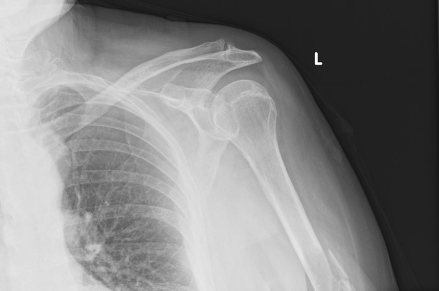
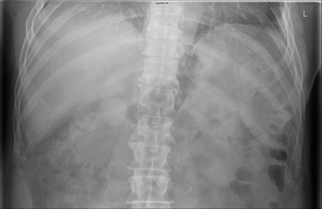
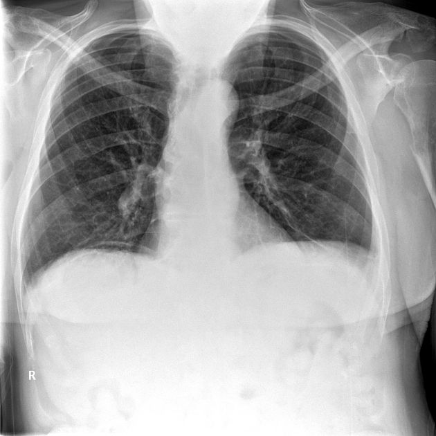
Edge of film errors, also known as corner of film errors, are a classical perceptual error in radiology where a pertinent finding, whether incidental or not, is at the margin or edge of the image.
It is now used for all modalities, in both a literal sense, i.e. actually at the edge of the image, or more metaphorically, perhaps just the last slice in an image series or only seen on a single MRI localizer image.
Under the Renfrew classification of errors in radiology, these errors are generally thought to be "type 4: under-reading", although it could also be argued that in some cases they are also "type 9: location", i.e. outside the area of interest 3.
References
- 1. Michael A. Bruno. Error and Uncertainty in Diagnostic Radiology. (2018) ISBN: 9780190665401
- 2. Jason N. Itri, Rafel R. Tappouni, Rachel O. McEachern, Arthur J. Pesch, Sohil H. Patel. Fundamentals of Diagnostic Error in Imaging. (2018) RadioGraphics. 38 (6): 1845-1865. doi:10.1148/rg.2018180021 - Pubmed
- 3. Bruno M, Walker E, Abujudeh H. Understanding and Confronting Our Mistakes: The Epidemiology of Error in Radiology and Strategies for Error Reduction. Radiographics. 2015;35(6):1668-1676. doi:10.1148/rg.2015150023
Incoming Links
Related articles: Errors and bias in diagnostic radiology
- errors in diagnostic radiology
-
cognitive bias in diagnostic radiology
- anchoring bias
- automation bias
- availability bias
- confirmation bias
- hindsight bias
- representativeness bias
- framing bias
- outcome bias
- length time bias
- lead time bias
- recall bias
- selection bias
- zebra retreat bias
Related articles: Terms used in radiology
- general
- ancillary
- Cinderella
- diagnosis of exclusion
- dilation vs dilatation
- epiphenomenon
- florid
- forme fruste
- gold standard
- heterogeneous vs heterogenous
- Hickam's dictum
- iatrogenic disease
- idiopathic
- in extremis
- natural history
- non-specific
- Occam's razor
- prodrome
- Saint's triad
- self-limiting
- sequela
- sine qua non
- status post
- subclinical disease
- syndrome
- radiology-specific
- pathology
- agenesis
- anlage
- aplasia
- apoptosis
- atresia
- atrophy
- cyst
- dehiscence
- diathesis
- diverticulum
- dyscrasia
- dysplasia
- exophytic
- fistula
- fluid collection
- granulation tissue
- hernia
- hyperplasia
- hypertrophy
- hypoplasia
- lamellated
- laminated
- malignancy
- metaplasia
- necrosis
- neoplasm
- phlegmon
- septum
- synechia
- trabecula
- CNS
- chest
- epidemiology
- gastrointestinal
- genetics
- musculoskeletal
- oncology


 Unable to process the form. Check for errors and try again.