
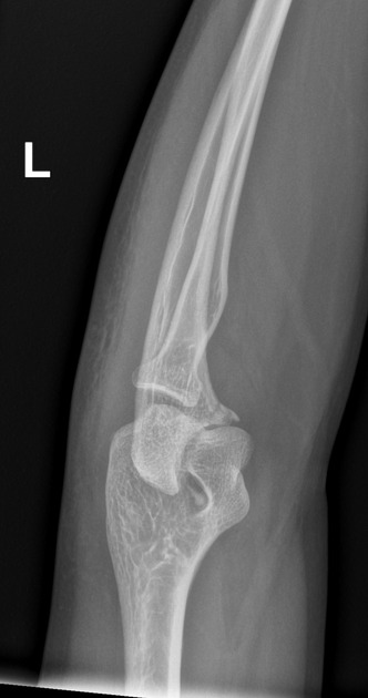
The elbow internal oblique view is a specialized projection, utilized to demonstrate both the coronoid process in profile and the olecranon process sitting within the olecranon fossa of the humerus. The affected limb is pronated.
On this page:
Indications
The medial oblique view is not a standard projection of the elbow, however helpful when occult fractures of the coronoid process are suspected.
Patient position
- the patient is seated alongside the table
- fully extended arm and forearm, in a pronated position
- ensure the anterior portion of the elbow is roughly 45 degrees from the IR
Technical factors
- anteroposterior medial oblique projection
-
centering point
- mid elbow joint
-
collimation
- superior to the distal third of the humerus
- inferior to include one-third of the proximal radius and ulna
- lateral to include the skin margin
- medial to include medial skin margin
-
orientation
- portrait
-
detector size
- 18 cm x 24 cm
-
exposure
- 50-60 kVp
- 2-5 mAs
-
SID
- 100 cm
-
grid
- no
Image technical evaluation
- the coronoid process should be seen in profile
- a clear visualization of the olecranon sitting in the olecranon fossa
- the radial head and proximal ulna are superimposed
Practical points
Keep in mind patient comfort when performing this examination.


 Unable to process the form. Check for errors and try again.