
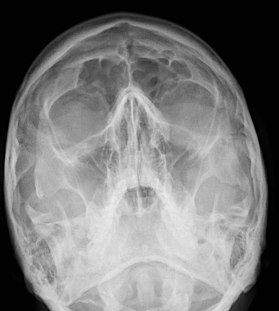
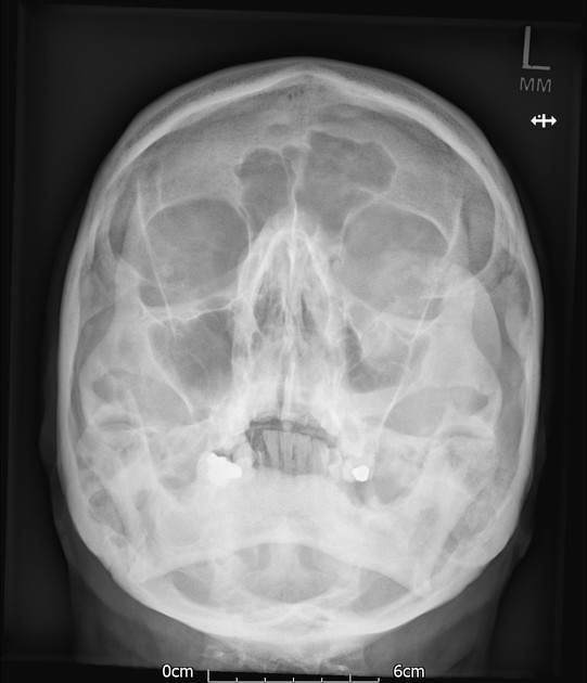
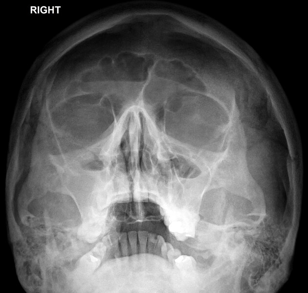
The occipitomental (OM) 4 or Waters view or parietoacanthial projection 2 is an angled PA radiograph of the skull, with the patient gazing slightly upwards.
On this page:
Indications
It can be used to assess for facial fractures, as well as for acute sinusitis. In general, radiographs of the skull and facial bones are rapidly becoming obsolete, being replaced by much more sensitive CT scans.
Patient position
the patient is erect facing the upright detector
the chin is raised until the mento-mandibular line (MML) is perpendicular to the receptor (orbitomeatal line (OML) will be 37° from receptor) 2
ensure patient's head is straight
Technical factors
posteroanterior projection
-
centering point
the beam is exiting at the acanthion 2
-
collimation
superior to the skin margins
inferior to include the most inferior aspects of the skull
lateral to include the skin margin
-
orientation
portrait
-
detector size
24 cm x 30 cm 2
-
exposure
75-80 kVp
20-25 mAs
-
SID
100 cm 2
-
grid
yes (this can vary departmentally)
Image technical evaluation
the petrous ridge should be inferior to the maxillary sinuses
assess for rotation via the assessments of the coronoid process symmetry
generally, the base of the mandible and the occiput will be superimposed
Practical points
learn your skull positioning lines, it makes for reading position guides a lot easier
guarantee that the patient is not "hunched" over when they are being examined, this can cause an artifact from the shoulders and the patient is more likely to be rotated; it is best to move the chair up close to the detector so they are sitting up straight for the image
use a side marker and regularly place in PA; skulls can get tricky with figuring out which side is which; many vendors tend to "flip" images to make them appear AP
History and etymology
This view was first described by Charles A Waters and C W Waldron, American radiologists, in 1915 3.


 Unable to process the form. Check for errors and try again.