
Forearm lateral view is one of two standard projections in the forearm series to assess the radius and ulna.
On this page:
Indications
This view allows for the assessment of suspected dislocations or fractures and localizing foreign bodies within the forearm.
Patient position
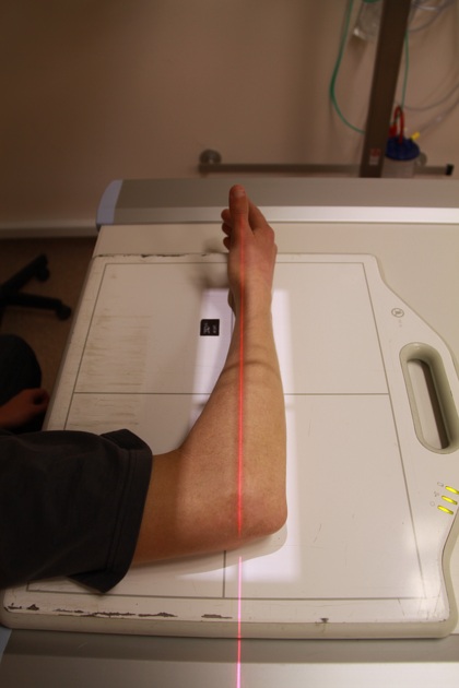
patient is seated alongside the table
elbow is flexed to 90 degrees, and the medial aspect of the wrist, forearm and elbow joint are placed in contact with the detector
shoulder, elbow and wrist should be in the same horizontal plane
Technical factors
lateral projection
-
centering point
mid forearm region
-
collimation
distal to the wrist joint
proximal to the elbow joint
-
orientation
portrait
-
detector size
24 cm x 30 cm
-
exposure
50-60 kVp
3-5 mAs
-
SID
100 cm
-
grid
no
Image technical evaluation
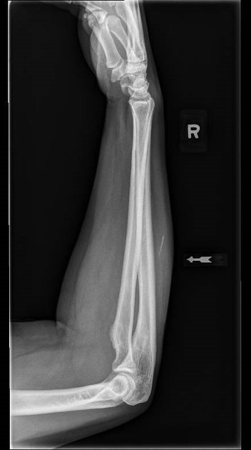
elbow is in a lateral position, as confirmation by the trochlea and capitulum being superimposed and the radial head being seen in profile
there should be superimposition of the distal radius and ulna indicating a lateral position
Practical points
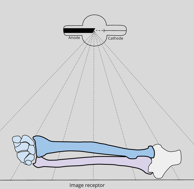
Contrary to popular belief the lateral forearm should not be considered a view to evaluating any occult injuries of the wrist joint and or elbow due to beam divergence. Beam divergence at the edges of the image should be acknowledged when assessing anatomy (see Figure 1) 2.


 Unable to process the form. Check for errors and try again.