
A variety of intracranial tumors exhibit different forms of calcification. Some lesions commonly show calcification while in some tumors, calcification is seen only in few number of cases. In this article these tumors are classified on the basis of frequency of calcification.
Commonly calcified tumors
Parenchymal
- oligodendroglioma: central or peripheral ribbon-like calcification
- desmoplastic infantile astrocytoma and ganglioglioma
- ganglioglioma/gangliocytoma
- cavernous hemangioma
- dysembryoplastic neuroepithelial tumor (DNET): more microscopic calcification
- atypical teratoid/rhabdoid tumor
- pilocytic astrocytoma
- calcifying pseudoneoplasms of the neuraxis (CAPNON) - more commonly extra-axial
Extra-axial
- meningioma
- chondrosarcoma of skull base: rings and arcs pattern
- chordoma
- intracranial dermoid
- intracranial teratoma: clump-like calcification
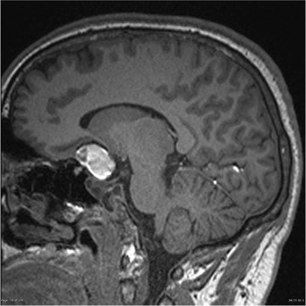
- adamantinomatous craniopharyngioma: stippled and peripheral craniopharyngioma (calcification is rare in papillary craniopharyngiomas)
- olfactory neuroblastoma
- tubulonodular pericallosal lipoma: peripheral bracket sign calcification
- calcifying pseudoneoplasms of the neuraxis (CAPNON) - can be intra-axial
Pineal region
- pineoblastoma/pineocytoma: exploded calcification
- pineal germinoma
Intraventricular
- central neurocytoma: punctuate calcification
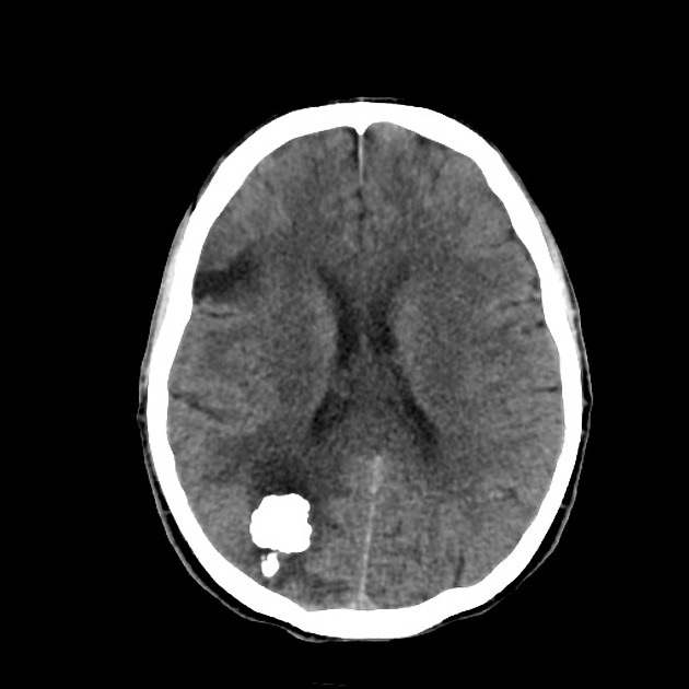
- choroid plexus papilloma: speckled calcification
- ependymoma: coarse calcification
- subependymoma


 Unable to process the form. Check for errors and try again.