
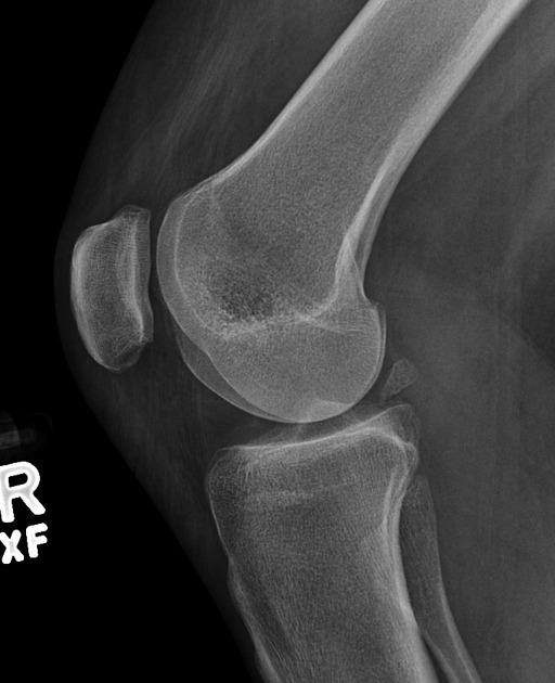
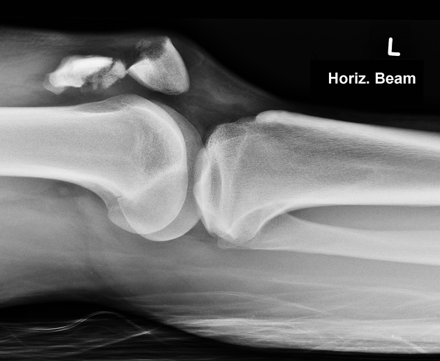
The lateral knee view is an orthogonal view of the AP view of the knee. The projection requires the patient to 'roll' onto the side of their knee, hence it is not an appropriate projection in trauma, in all suspected traumatic injuries of the knee, the horizontal beam lateral method should be utilized.
On this page:
Indications
This is often performed on patients with suspected arthritis, it is an orthogonal view of the AP projection and demonstrates the spaces of the knee joint, yet sacrifices any assessment of fluid levels.
Patient position
the patient is lateral recumbent with the knee of interest closest to the table and the other lower limb rolled anteriorly
affected knee is flexed slightly ≈ 30° (to the best of patient's ability); anything more than 30° is less than ideal as the patella will move inferiorly and the soft tissues will begin to compress
Technical factors
medial-lateral projection
-
centering point
center to the knee joint 1.5-2.0 cm distal to the apex of the patella or at the tibial tuberosity if the patella is affected by certain injury patterns
-
collimation
superior to include the distal femur
inferior to include the proximal tibia/fibula
anteroposteriorly to include skin margin
-
orientation
landscape
-
detector size
35 cm x 43 cm
-
exposure
60-70 kVp
7-10 mAs
-
SID
100 cm
-
grid
no
Image technical evaluation
A true lateral projection will have the following characteristics:
superimposition of the medial and lateral condyles of the distal femur
an open patellofemoral joint space
slight superimposition of the fibular head with the tibia
Practical points
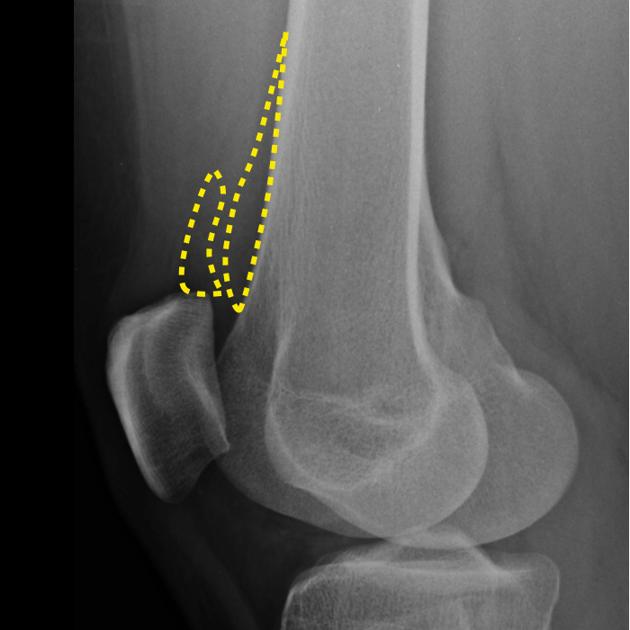
The distal femoral condyles have distinct features that can be used for differentiation and hence positional errors that can be corrected:
the medial condyle has a medial adductor tubercle, located superior to the medial epicondyle, a bony protuberance that acts as the attachment point the adductor minimus and the hamstrings part of the adductor magnus
the lateral condyle has the condylopatellar sulcus also known as the lateral notch, a groove in the lateral femoral condyle. The easy way to remember is femoral is flat
To superimpose the medial and lateral femoral condyles, use a:
cephalic tilt of 4 - 7°
adduct the patient's leg to the mid-sagittal plane by 4 - 7°
Note: Patients with a total knee replacement generally do not require a cephalic tilt/adduction
Correcting rotational errors
-
medial adductor tubercle is posterior to the lateral condyle
rotate the knee externally to bring it anterior
-
medial adductor tubercle is anterior to the lateral condyle
rotate the knee internally to bring it posteriorly
Abduction and adduction
-
medial condyle is inferior to the lateral condyle
perform adduction
-
medial condyle is superior to the lateral condyle
perform abduction
For an interactive case exploring these concepts see here


 Unable to process the form. Check for errors and try again.