
The knee series is a set of radiographs taken to investigate knee joint pathology, often in the context of trauma. It usually comprises an AP and lateral projection, although other non-standard, modified projections can be used for specific indications.
See also knee radiograph (an approach).
Indications
Knee radiographs are indicated for a variety of settings including 1,2:
trauma
bony tenderness at the head of the fibula
isolated patella tenderness
patient unable to flex the knee to 90 degrees
if the patient is unable to bear weight
suspected osteoarthritis
detecting joint effusions
infection
Projections
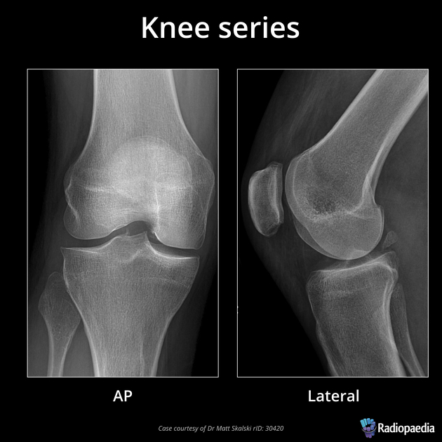
Standard projections
-
demonstrates the distal femur, proximal tibia/fibula and the patella in the AP position
ideal projection to assess the tibial plateau and tibiofemoral alignment
-
orthogonal projection to the AP, routinely done in trauma horizontal beam to better demonstrate any joint effusions
ideal projection to assess lipohemarthrosis
used to examine the location of the patella and the patency of the patella tendon
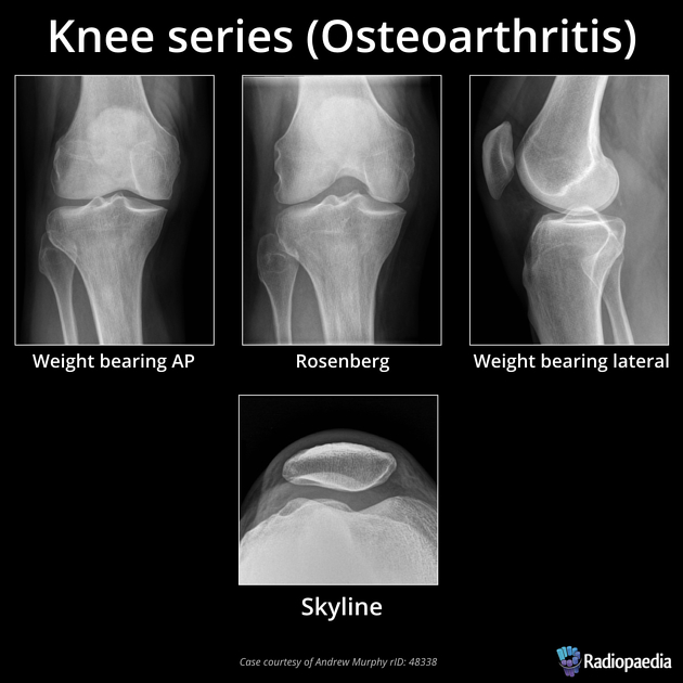
Additional projections
-
often performed on bed-bound patients with suspected arthritis
orthogonal view of the AP projection
demonstrate the joint space, yet sacrifices any assessment of fluid levels
-
superior-inferior projection of the patella; this is an ideal projection for patients that are better suited in the supine position
-
inferior-superior projection of the patella; this projection is best suited to patients able to maintain a semi-recumbent position on the examination table
-
often used in the context of orthopedic appointments to obtain images of the knees in their natural anatomical position
-
view utilized to demonstrate intercondylar space, often used for OA and suspected tibial plateau fractures
-
weight-bearing projection used to assess joint space-related pathology such as osteoarthritis
-
two views (internal and external) better demonstrating the knee joint in the absence of CT


 Unable to process the form. Check for errors and try again.