
Left upper lobe collapse
Updates to Article Attributes
Left upper lobe collapse has distinctive features but can be challenging to identify on chest radiographs by the uninitiated. Hence it is a classic chest radiograph case for radiology fellowships exams.
For a general discussion refer to the article on lobar collapse.
Radiographic features
Plain radiograph
The left upper lobe collapses anteriorly becoming a thin sheet of tissue apposed to the anterior chest wall and appears as a hazy or veiling opacity extending out from the left hilum and fading out inferiorly 1. It thus reverses the normal slight increase in radiographic density seen as you move down the lung (due to the increased thickness of the chest soft tissues). This is known as veiling density and can be subtle.
Parts of the normal cardiomediastinal contour may also be obscured where the left upper lobe, particularly the lingular segments, abuts the left heart border. The anterior parts of the aortic arch are also often obscured.
In some cases the hyperexpanded superior segment of the left lower lobe insinuates itself between the left upper lobe and the superior mediastinum, sharply silhouetting the aortic arch and resulting in a sickle-shaped lucency medially. This is known as the luftsichel sign 1.
If the collapse is not great enough, the hyperinflated superior segment of the left lower lobe may cause an apparent apical cap, which can be mistaken for pleural fluid or thickening or even a Pancoast tumour 2.
The left hilum is drawn upwards, resulting in an almost horizontal course of the left main bronchus and vertical course of the left lower lobe bronchus 1.
Non-specific signs indicating left sided atelectasis will also be present, including:
- elevation of the hemidiaphragm
- 'peaked' or 'tented' hemidiaphragm: juxtaphrenic peak sign
- crowding of the left ribs
- shift of the mediastinum to the left
- posterior and left lateral rotation of the heart
On lateral projections the left lower lobe collapse causes increase retrosternal density and the oblique fissure displaced anteriorly. There is associated anterior displaced of the left hilum.
CT
- triangular opacification in axial images, thinner at the hilum 2
- the oblique fissure rotates anteromedially 2
Differential diagnosis
- left upper lobe consolidation with the absence of signs of volume loss
See alsoRelated articles
See also
-<a href="/articles/left-upper-lobe-consolidation">left upper lobe consolidation</a> with the absence of signs of volume loss</li></ul><h4>See also</h4><ul>- +<a href="/articles/left-upper-lobe-consolidation">left upper lobe consolidation</a> with the absence of signs of volume loss</li></ul><h4>Related articles</h4><ul>
-<li><a href="/articles/lobar-lung-collapse">lobar collapse</a></li>-</ul>- +<li>other <a href="/articles/lobar-lung-collapse">lobar collapses</a> (<a href="/articles/lobar-collapse-summary">collapse summary article</a>)<ul>
- +<li><a href="/articles/right-upper-lobe-collapse">right upper lobe collapse</a></li>
- +<li><a href="/articles/right-middle-lobe-collapse">right middle lobe collapse</a></li>
- +<li><a href="/articles/right-lower-lobe-collapse">right lower lobe collapse</a></li>
- +<li><a href="/articles/left-lower-lobe-collapse">left lower lobe collapse</a></li>
- +</ul>
- +</li>
- +</ul><h4>See also</h4><ul><li><a href="/articles/left-upper-lobe-collapse-in-the-exam">left upper lobe collapse in an exam setting</a></li></ul>
Image 1 Annotated image (Frontal) ( create )

Image 2 Annotated image (Lateral) ( create )

Image 3 Annotated image (Frontal) ( update )

Image 4 Annotated image (Lateral) ( update )

Image 5 X-ray (Lateral) ( update )

Image 6 X-ray (Frontal) ( update )

Image 7 X-ray (Frontal) ( update )

Image 8 X-ray (Frontal) ( update )

Image 9 X-ray (Frontal) ( update )

Image 10 X-ray (Frontal) ( update )

Image 11 X-ray (Frontal) ( update )
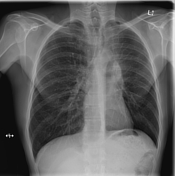
Image 12 X-ray (Frontal) ( update )

Image 14 X-ray (Frontal) ( update )
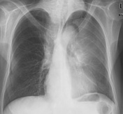
Image 15 X-ray (Frontal) ( update )

Image 16 X-ray (Frontal) ( update )
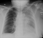
Image 17 CT (lung window) ( update )


 Unable to process the form. Check for errors and try again.