
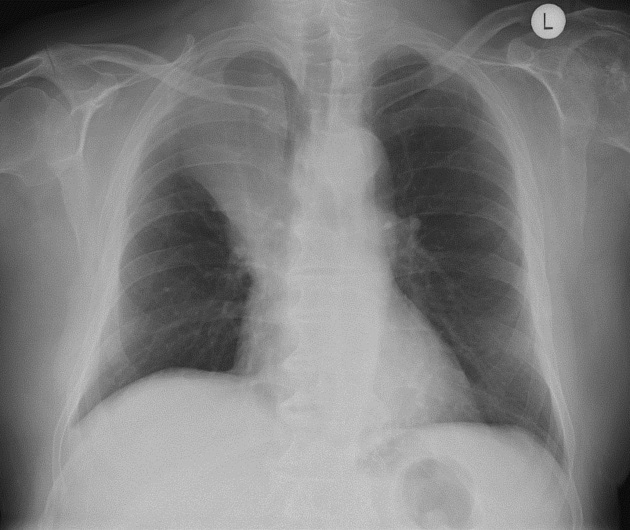
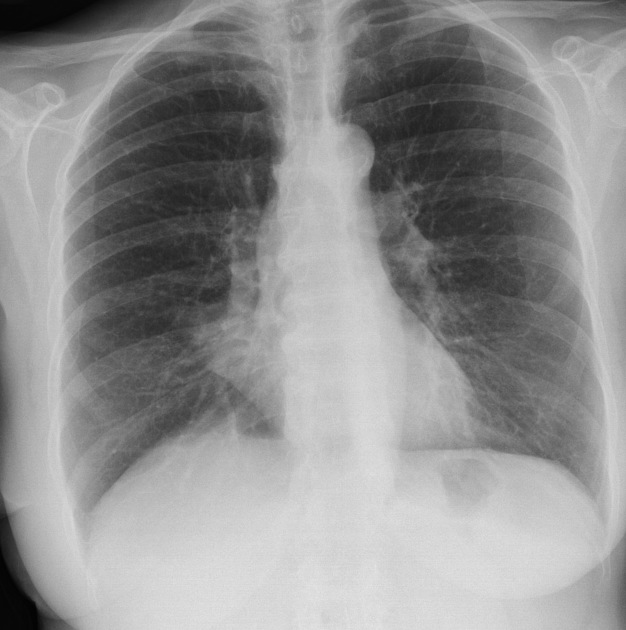
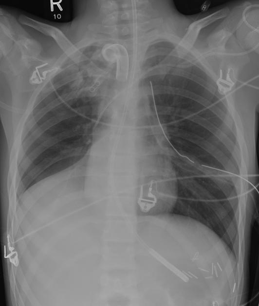
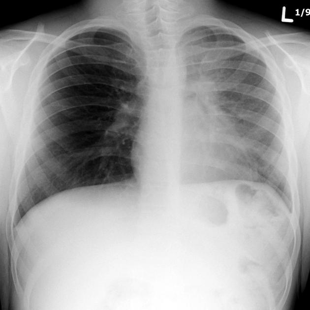
Lobar lung collapse
Citation, DOI, disclosures and article data
At the time the article was created Frank Gaillard had no recorded disclosures.
View Frank Gaillard's current disclosuresAt the time the article was last revised Raymond Chieng had no financial relationships to ineligible companies to disclose.
View Raymond Chieng's current disclosures- Lobar atelectasis
- Lobar lung atelectasis
- Lobar collapse
- Lobal volume loss
- Lobar volume loss
Lobar collapse refers to the collapse of an entire lobe of the lung. It is a subtype of atelectasis. Individual lobes of the lung may collapse due to obstruction of the supplying bronchus 12.
Pathology
Most often collapse of most or all of a lobe is secondary to bronchial obstruction causing resorptive atelectasis.
Etiology
-
luminal
aspirated foreign material
endobronchial mass
-
mural
-
extrinsic
compression by adjacent mass
Radiographic features
There are several classical rules that a lobar collapse follows 9:
bowing or displacement of a fissure/s occurs towards the collapsing lobe
a significant amount of volume loss is required to cause air space opacification
the collapsed lobe is triangular or pyramidal in shape, with the apex pointing to the hilum
-
the collapsed lung peripherally maintains contact with the costal parietal pleura, except:
in RML collapse where the lobe collapses adjacent to the mediastinum
in the presence of pleural effusion
in the presence of pneumothorax
Several factors may influence the typical appearance of lobar collapse, including pre-existing lung disease, amount of volume loss, concomitant consolidation, pleural effusion or the presence of pneumothorax.
Plain radiograph
Generally, there is pulmonary air space opacification but the appearance on chest x-ray varies according to the lobe involved and are discussed separately:
Some features, however, are generic markers of volume loss and are helpful in directing one's attention to the collapse, as well as enabling distinction from opacification of the lobe without collapse (i.e. consolidation e.g. lobar pneumonia). These features include 5,9:
-
direct signs
displacement of fissures
crowding of pulmonary vessels
-
indirect signs
elevation of the ipsilateral hemidiaphragm
crowding of the ipsilateral ribs
shift of the mediastinum towards the side of atelectasis
compensatory hyperinflation of normal lobes
hilar displacement towards the collapse
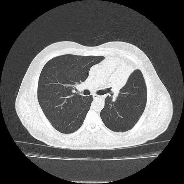
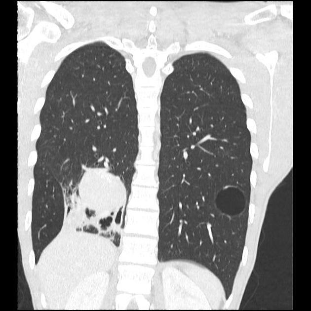
CT
Lobar collapse is usually trivially easy to identify on CT, but identification of the cause is not always easy, as the collapsed lung can make identification of an obstructing lesion difficult. The density of the collapsed lobe is high post contrast administration.
References
- 1. Proto AV, Tocino I. Radiographic manifestations of lobar collapse. Semin Roentgenol. 1980;15 (2): 117-73. - Pubmed citation
- 2. Robbins LL, Clayton HH. The roentgen appearance of lobar and segmental collapse of the lung. VI. Collapse of the upper lobes. Radiology 1945; 45:347-355.
- 3. Lubert M, Krause GR. Further observations on lobar collapse. Radiol Clin North Am 1963; 1:331-346.
- 4. Lee SK, Ahn JM, Im J, Muller NL. Lobar atelectasis: typical and atypical radiographic and CT findings. Postgrad Radiol 1995; 15:203-217.
- 5. Collins J, Stern EJ. Chest radiology, the essentials. Lippincott Williams & Wilkins. (2007) ISBN:0781763142. Read it at Google Books - Find it at Amazon
- 6. Woodring JH, Reed JC. Radiographic manifestations of lobar atelectasis. J Thorac Imaging. 1996;11 (2): 109-44. - Pubmed citation
- 7. Ashizawa K, Hayashi K, Aso N et-al. Lobar atelectasis: diagnostic pitfalls on chest radiography. Br J Radiol. 2001;74 (877): 89-97. Br J Radiol (citation) - Pubmed citation
- 8. Lee KS, Logan PM, Primack SL et-al. Combined lobar atelectasis of the right lung: imaging findings. AJR Am J Roentgenol. 1994;163 (1): 43-7. AJR Am J Roentgenol (citation) - Pubmed citation
- 9. W. Richard Webb, Charles B. Higgins. Thoracic Imaging. (2010) ISBN: 9781605479767
- 10. Proto AV. Lobar collapse: basic concepts. (1996) European journal of radiology. 23 (1): 9-22. Pubmed
- 11. Woodring JH, Reed JC. Types and mechanisms of pulmonary atelectasis. (1996) Journal of thoracic imaging. 11 (2): 92-108. Pubmed
- 12. Bankier A, MacMahon H, Colby T et al. Fleischner Society: Glossary of Terms for Thoracic Imaging. Radiology. 2024;310(2):e232558. doi:10.1148/radiol.232558 - Pubmed
Incoming Links
- Lobar pneumonia
- Resorptive (obstructive) atelectasis
- Chronic eosinophilic pneumonia
- Kawasaki disease
- Right middle lobe collapse
- V/Q scan
- Right upper lobe collapse
- Left lower lobe collapse
- Sickle cell disease (acute chest syndrome)
- Tracheal bifurcation angle
- Shifting granuloma sign
- Left upper lobe collapse
- Usual interstitial pneumonia
- Juxtaphrenic peak sign
- Lung atelectasis
- Pulmonary mycobacterium abscessus infection
- Unilateral hypertransradiant hemithorax
- Mediastinal widening (differential)
- Acute aspiration pneumonitis
- Crack lung
- Lobar collapse - secondary to endobronchial valve insertion
- Monobronchial intubation.
- Lobar pneumonia
- Hydropneumothorax
- Non-small cell lung carcinoma
- Congenital diaphragmatic hernia (Bochdalek hernia)
- Left lower lobe collapse
- Bronchial carcinoid tumour
- Right lower lobe collapse (foreign body)
- Left upper lobe collapse
- Left lower lobe collapse
- Loss of AP window secondary to pleural tumour
- Left upper lobe collapse due to lung cancer
- Right lower lobe collapse
- Right upper lobe collapse
- Left upper lobe collapse
- Right upper lobe collapse
- Left lower lobe collapse
- Acute myocardial infarction in CT
- Persistent pneumothorax due to right lung collapse and bronchial obstruction from haematoma
Related articles: Airspace opacification
- airspace opacification
- differential diagnoses of airspace opacification[+][+]
- lobar consolidation[+][+]
-
atelectasis[+][+]
- mechanism-based
- morphology-based
- lobar lung collapse
Related articles: Chest
- imaging techniques
-
chest radiograph
- radiography[+][+]
-
approach
- ABCDE
- ABCDEFGHI
- congenital heart disease
- medical devices in the thorax
- common lines and tubes[+][+]
- nasogastric tubes
- endotracheal tubes
- central venous catheters
- esophageal temperature probe
- tracheostomy tube
- pleural catheters
- cardiac conduction devices
- prosthetic heart valve
- review areas
-
airspace opacification
- differential diagnoses of airspace opacification[+][+]
- lobar consolidation[+][+]
-
atelectasis[+][+]
- mechanism-based
- morphology-based
- lobar lung collapse
- chest x-ray in the exam setting[+][+]
- cardiomediastinal contour[+][+]
- chest radiograph zones[+][+]
- tracheal air column[+][+]
- fissures[+][+]
- normal chest x-ray appearance of the diaphragm[+][+]
- nipple shadow[+][+]
-
lines and stripes[+][+]
- anterior junction line
- posterior junction line
- right paratracheal stripe
- left paratracheal stripe
- posterior tracheal stripe/tracheo-esophageal stripe
- posterior wall of bronchus intermedius
- right paraspinal line
- left paraspinal line
- aortic-pulmonary stripe
- aortopulmonary window
- azygo-esophageal recess
- spaces[+][+]
- signs[+][+]
- air bronchogram
- big rib sign
- Chang sign
- Chen sign
- coin lesion
- continuous diaphragm sign
- dense hilum sign
- double contour sign
- egg-on-a-string sign
- extrapleural sign
- finger in glove sign
- flat waist sign
- Fleischner sign
- ginkgo leaf sign
- Golden S sign
- Hampton hump
- haystack sign
- hilum convergence sign
- hilum overlay sign
- Hoffman-Rigler sign
- holly leaf sign
- incomplete border sign
- juxtaphrenic peak sign
- Kirklin sign
- medial stripe sign
- melting ice cube sign
- more black sign
- Naclerio V sign
- Palla sign
- pericardial fat tag sign
- Shmoo sign
- silhouette sign
- snowman sign
- spinnaker sign
- steeple sign
- straight left heart border sign
- third mogul sign
- tram-track sign
- walking man sign
- water bottle sign
- wave sign
- Westermark sign
- HRCT[+][+]
-
chest radiograph
- airways[+][+]
- bronchitis
- small airways disease
-
bronchiectasis
- broncho-arterial ratio
- related conditions
- differentials by distribution
- narrowing
-
tracheal stenosis
- diffuse tracheal narrowing (differential)
-
bronchial stenosis
- diffuse airway narrowing (differential)
-
tracheal stenosis
- diverticula
- pulmonary edema[+][+]
-
interstitial lung disease (ILD)[+][+]
- Anti-Jo-1 antibody-positive interstitial lung disease
- drug-induced interstitial lung disease
-
hypersensitivity pneumonitis
- acute hypersensitivity pneumonitis
- subacute hypersensitivity pneumonitis
- chronic hypersensitivity pneumonitis
- etiology
- bird fancier's lung: pigeon fancier's lung
- farmer's lung
- cheese workers' lung
- bagassosis
- mushroom worker’s lung
- malt worker’s lung
- maple bark disease
- hot tub lung
- wine maker’s lung
- woodsman’s disease
- thatched roof lung
- tobacco grower’s lung
- potato riddler’s lung
- summer-type pneumonitis
- dry rot lung
- machine operator’s lung
- humidifier lung
- shower curtain disease
- furrier’s lung
- miller’s lung
- lycoperdonosis
- saxophone lung
-
idiopathic interstitial pneumonia (mnemonic)
- acute interstitial pneumonia (AIP)
- cryptogenic organizing pneumonia (COP)
- desquamative interstitial pneumonia (DIP)
- non-specific interstitial pneumonia (NSIP)
- idiopathic pleuroparenchymal fibroelastosis
- lymphoid interstitial pneumonia (LIP)
- respiratory bronchiolitis–associated interstitial lung disease (RB-ILD)
- usual interstitial pneumonia / idiopathic pulmonary fibrosis (UIP/IPF)
-
pneumoconioses
- fibrotic
- non-fibrotic
-
lung cancer[+][+]
-
non-small-cell lung cancer
-
adenocarcinoma
- pre-invasive tumors
- minimally invasive tumors
- invasive tumors
- variants of invasive carcinoma
- described imaging features
- adenosquamous carcinoma
- large cell carcinoma
- primary sarcomatoid carcinoma of the lung
- squamous cell carcinoma
- salivary gland-type tumors
-
adenocarcinoma
- pulmonary neuroendocrine tumors
- preinvasive lesions
-
lung cancer invasion patterns
- tumor spread through air spaces (STAS)
- presence of non-lepidic patterns such as acinar, papillary, solid, or micropapillary
- myofibroblastic stroma associated with invasive tumor cells
- pleural invasion
- vascular invasion
- tumors by location
- benign neoplasms
- pulmonary metastases
- lung cancer screening
- lung cancer staging
-
non-small-cell lung cancer


 Unable to process the form. Check for errors and try again.