
The long axial hindfoot alignment view is a specialized, weight-bearing radiographic view that examines the hindfoot alignment as part of a foot and ankle instability investigation.
The long axial view requires no equipment and has higher inter-observer reliability compared to the standard hindfoot alignment view when measuring angular hindfoot alignment 1,2.
On this page:
Indications
Suspected varus or vagus malalignment 1,2.
Patient position
- the patient stands on a flat detector on the floor
- alternatively, on a radiolucent stand with the upright detector in the horizontal position under the detector
- patient distributes weight evenly across both feet with the foot in question central to the detector
- the foot is in a neutral position
Technical factors
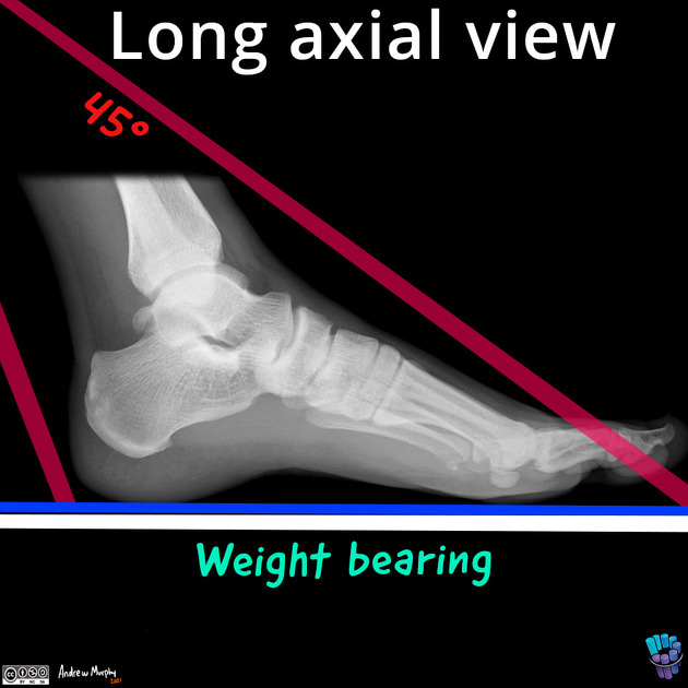
- posterior-anterior, axial projection
-
centering point
- the central beam is angled 45° toward the floor
- centered at the midpoint of the lateral and medial malleoli
-
collimation
- at least half the tibia and the calcaneum need to be included
-
orientation
- portrait
- detector size
- 18 cm x 24 cm
-
exposure
- 50-60 kVp
- 3-6 mAs
-
SID
- 100 cm
-
grid
- no
Image technical evaluation
- clear identification of the most distal portion of the calcaneus
- clear identification of the anatomical axis of the tibia
Practical points
It is important not to collimate tightly on this projection as the anatomical axis of the tibia must be established to calculate the hindfoot alignment. This is calculated via the distance between the anatomical axis of the tibia and the lowest part of the calcaneus (normal sitting at a mean value of 3.2mm) 1.


 Unable to process the form. Check for errors and try again.