
Lower limb radiography
Citation, DOI, disclosures and article data
Citation:
Hacking C, Sharma R, Walizai T, et al. Lower limb radiography. Reference article, Radiopaedia.org (Accessed on 09 Mar 2025) https://doi.org/10.53347/rID-39131
rID:
39131
Article created:
Disclosures:
At the time the article was created Craig Hacking had no recorded disclosures.
View Craig Hacking's current disclosures
Last revised:
Disclosures:
At the time the article was last revised Rohit Sharma had no financial relationships to ineligible companies to disclose.
View Rohit Sharma's current disclosures
Revisions:
6 times, by
5 contributors -
see full revision history and disclosures
Systems:
Sections:
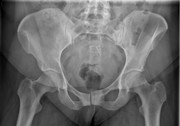
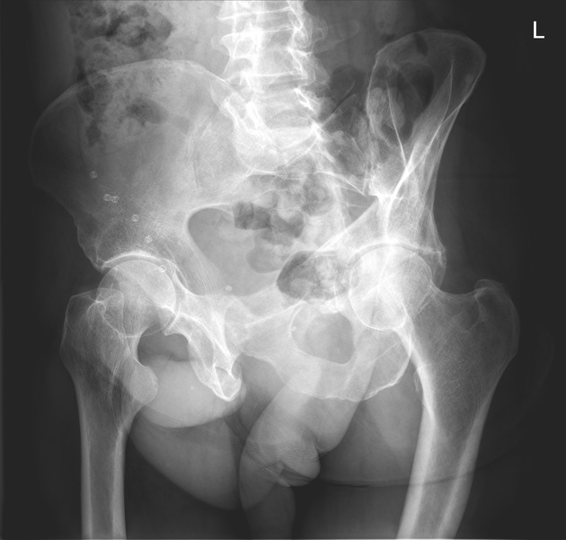
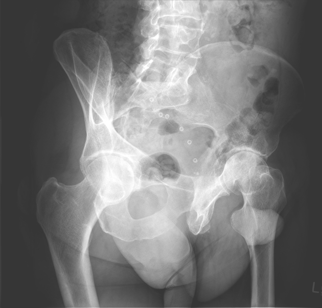
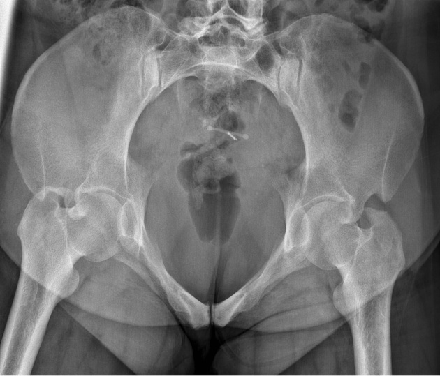
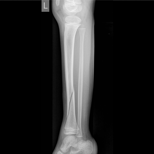
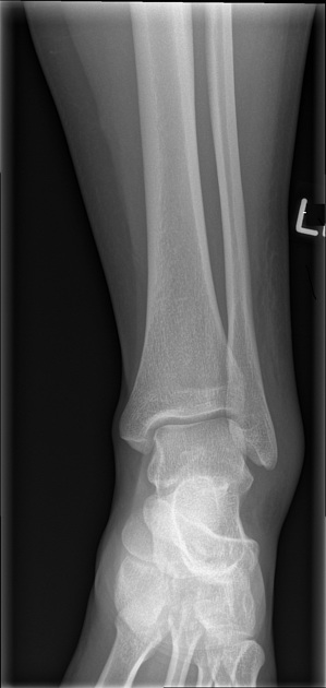
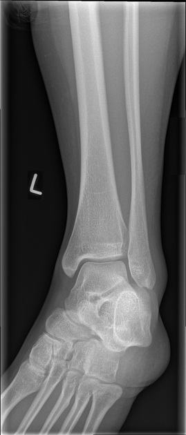
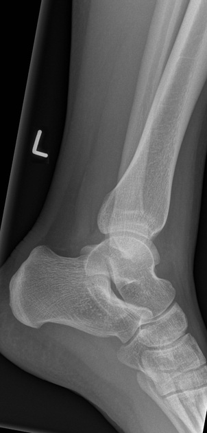
Lower limb radiography is the radiological investigation of the pelvis, hip joint, femur, knee joint, tibia, fibula, ankle joint, tarsal bones of the foot and metatarsals. It is often utilized in the context of trauma to rule out fractures and dislocations. Sometimes it can be used in skeletal surveys as well.
Related articles: Radiographs (adult)
- general radiography (adult)
- radiographic positioning terminology[+][+]
-
systematic radiographic technical evaluation (mnemonic)
- portable radiography
- chest radiography[+][+]
- abdominal radiography[+][+]
-
upper limb radiography[+][+]
-
shoulder girdle radiography
- scapula series
-
shoulder series
- shoulder (AP view)
- shoulder (internal rotation view)
- shoulder (external rotation view)
- shoulder (superior-inferior axial view)
- shoulder (inferior-superior axial)
- shoulder (West Point view)
- shoulder (Velpeau view)
- shoulder (modified trauma axial view)
- shoulder (supine lateral view)
- shoulder (modified transthoracic supine lateral)
- shoulder (lateral scapula view)
- shoulder (AP glenoid view)
- shoulder (Garth view)
- shoulder (outlet view)
- shoulder (Stryker notch view)
- acromioclavicular joint series
-
clavicle series
- clavicle (AP view)
- clavicle (AP cephalic view)
- clavicle (oblique view)
- sternoclavicular joint series
- arm and forearm radiography
- wrist and hand radiography
- wrist series
- scaphoid series
- hand series
- thumb series
- fingers series
- rheumatology hands series
- bone age (radiograph)
-
shoulder girdle radiography
-
lower limb radiography
- pelvic girdle radiography
- thigh and leg radiography
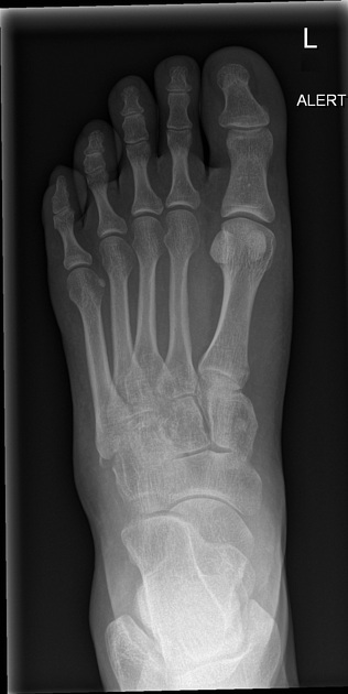
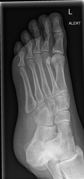
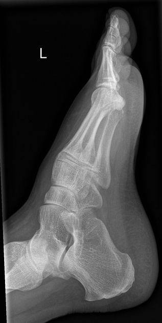
- ankle and foot radiography
- skull radiography[+][+]
-
paranasal sinus and facial bone radiography[+][+]
- facial bones
- mandible
- nasal bone
- zygomatic arches
- paranasal sinuses
- temporal bones
- dental radiography[+][+]
- orthopantomography
- temporomandibular joints
- temporomandibular joint (AP axial view)
- temporomandibular joint (axiolateral oblique view)
-
spinal radiography[+][+]
- cervical spine series
-
thoracic spine series
- thoracic spine (AP view)
- thoracic spine (lateral view)
- thoracic spine (oblique view)
- lumbar spine series
- sacrococcygeal radiography


 Unable to process the form. Check for errors and try again.