
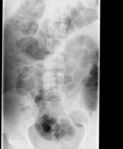
The lumbar spine anteroposterior or posteroanterior view images the lumbar spine in its anatomical position. The lumbar spine generally consists of five vertebrae (see: lumbosacral transitional vertebra).
On this page:
Indications
This projection is utilized in many imaging contexts including trauma, postoperatively, and for chronic conditions. Ideally, spinal imaging should be taken erect in the non-trauma setting to give a functional overview of the lumbar spine. Otherwise, patients with a suspected spinal injury must remain in the supine position without any movement.
Patient position
the patient is erect or supine, depending on clinical history
in the supine projection, hands are placed by the patient's side
-
if performing erect, position the patient in the PA position; this has numerous advantages including reduced dose to the gonadal region and utilization of beam divergence; arms can be placed by the side, or the handlebars of the erect Bucky can be held for patient stability
the weight bearing PA view can be called the Ferguson technique
Technical factors
anteroposterior projection
suspended expiration (for a uniform density)
-
centering point
the level of the iliac crests at the MSP
the central ray is perpendicular to the image receptor
-
collimation
superiorly to include the T12/L1 junction
inferior to include the sacral region
lateral to include the transverse processes and sacroiliac joints
-
orientation
portrait
-
detector size
35 cm x 43 cm
-
exposure
70-80 kVp
40-60 mAs
-
SID
110 cm
-
grid
yes (ensure the correct grid is selected if using focused grids)
Image technical evaluation
the entire lumbar spine should be visible, with demonstration of T11/T12 superiorly and the sacrum inferiorly.
no patient rotation as evident by central spinous processes and the symmetrical appearance of the sacroiliac joints and iliac wings
intervertebral joints are visualized
adequate image penetration and image contrast is evident by clear visualization of lumbar vertebral bodies, pedicles, and facet joints, with both trabecular and cortical bone demonstrated
Practical points
the three column concept of thoracolumbar spinal fractures is of particular importance when assessing this image for pathology
take particular care when imaging patient on a trauma trolley that the image receptor is aligned to the central ray to prevent anatomy exclusion and grid cut-off
ideally, the transverse processes should be visible, although demonstration is often obscured by overlying bowel gas; radiographers should ensure over exposure is not a factor contributing to the poor visualization which could mask a transverse process fracture
when imaging in a supine position, a triangular cushion can be placed under flexed knees to reduce lumbar lordosis, and thus aiding to open the intervertebral joints


 Unable to process the form. Check for errors and try again.