
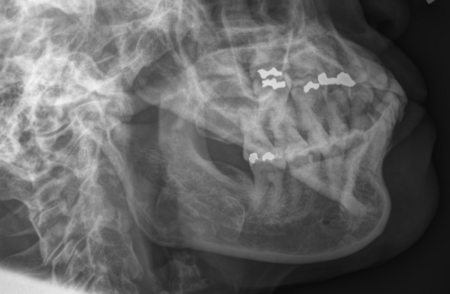
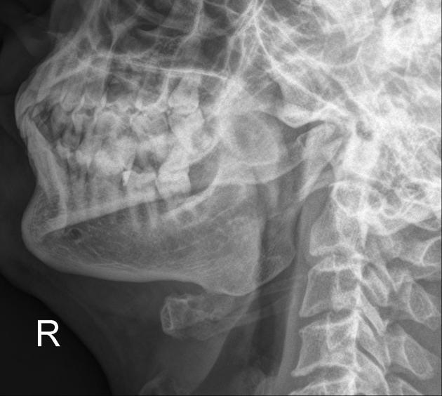
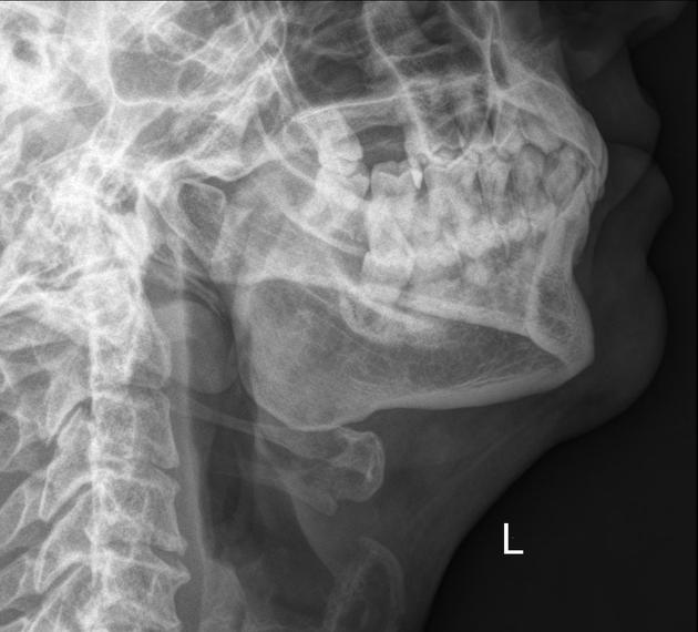
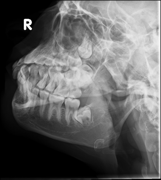
The axiolateral oblique mandible view allows for visualization of the mandibular body, mandibular ramus, condylar process and mentum.
On this page:
Indications
This projection is useful in identifying structural changes and displaced fractures of the mandible in a trauma setting, and in neoplastic or inflammatory changes. Given that this view is performed bilaterally, it allows for comparison of both sides of the mandible too.
Patient position
the patient is seated upright with the side of interest closest to the detector
-
the head is first placed in a true lateral position
interpupillary line (IPL) perpendicular and midsagittal plane (MSP) parallel to the detector
then, the neck is sufficiently extended to prevent superimposing the mandibular rami over the cervical spine
-
the vertex (top of the head) is lastly tilted towards the detector to
demonstrate the region of the mandible of interest
prevent superimposing the opposite side
Technical factors
left and right axiolateral oblique
-
centering point
central ray 25-30º cephalic, beam to exit at mandibular region of interest
-
collimation
no more than 10 x 10 cm with mandible of interest in the middle of the image
-
orientation
portrait
-
detector size
18 cm x 24 cm
-
exposure
70-75 kVp
16-25 mAs
-
SID
100 cm
-
grid
yes
Image technical evaluation
the ramus of interest is shown with no superimposition of the opposite mandible
the mandibular ramus is not superimposed over the cervical spine
Practical points
set up the x-ray tube and detector in advance; due to neck extension and tilt, patients may struggle to remain in the particular position for long, hence reducing image stability


 Unable to process the form. Check for errors and try again.