
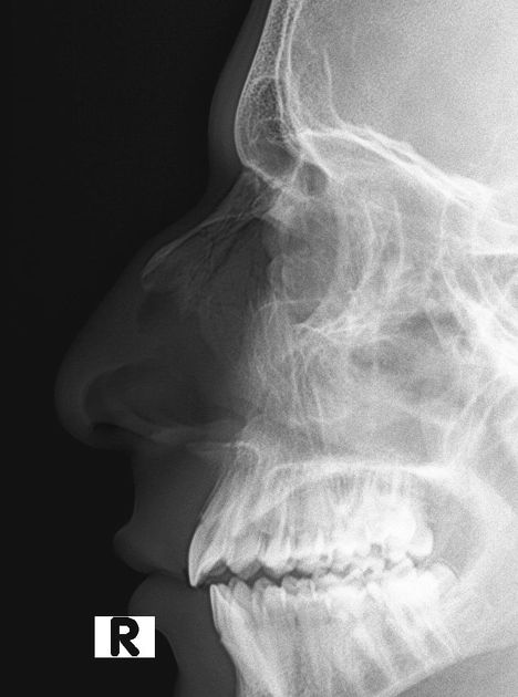
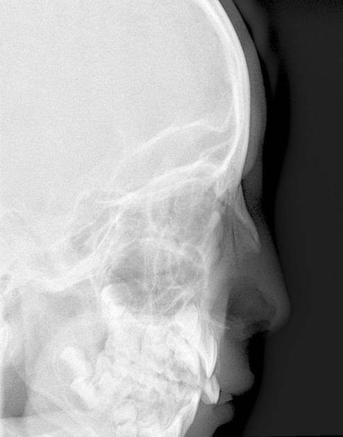
The lateral nasal bones view is a non-angled lateral radiograph showcasing two small oblong nasal bones situated side by side, together forming the nasal ridge.
On this page:
Indications
This view is often primarily used in assessing various nasal bone fractures in the trauma setting. Depending on the department, this view may be done bilaterally (for comparison of affected versus unaffected side) or unilaterally (only affected side).
Patient position
rest the lateral aspect of the patient's head (with the side of interest) against the image detector
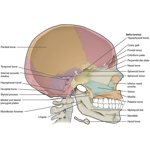
adjust the head into a true lateral position, with the midsagittal plane (Figure 1) parallel to the image detector
for patient's comfort, adjust the patient's body into an oblique position
Technical factors
lateral projection
-
centering point
1.25 cm inferior to nasion
-
collimation
within 5 cm of the nasal bones on all sides 1
-
orientation
for unilateral side: portrait
for bilateral side: landscape
-
detector size
24 cm x 18 cm
-
exposure
60-70 kVp
5-10 mAs
-
SID
100 cm
-
grid
no
Image technical evaluation
nasal bones with soft tissue nasal structure, frontonasal suture (superior) and anterior nasal spine should be demonstrated
nasal bones should be seen with no rotation
Practical points
remove glasses and nose piercings to avoid artifact obscuring important pathology
-
this view should not replace a lateral facial bones x-ray
as this projection is often requested together with a facial bones series 3, there may be the temptation to combine the request and only image a lateral facial bones x-ray as per ALARA. However, nasal bones and their soft tissue nasal structures are less dense compared to the denser facial bones and should hence be imaged separately to prevent over exposure of the less dense region


 Unable to process the form. Check for errors and try again.