
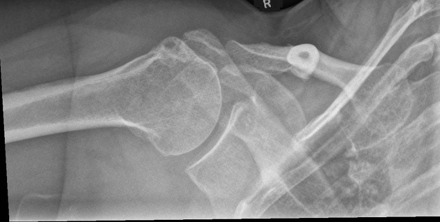
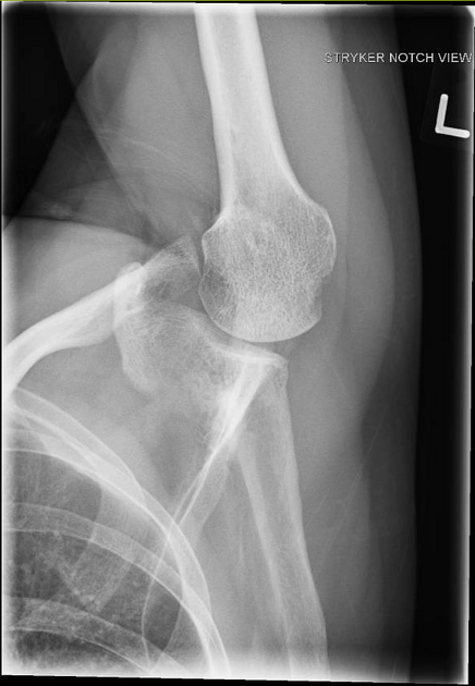
The Stryker notch view is a specialized projection of the shoulder, aimed at assessing the posterior humerus.
On this page:
Indications
The Stryker notch view can be used post anterior glenohumeral dislocation, assessing for Hill-Sachs defects 1.
Patient position
- the patient is preferably erect
- the mid-coronal plane of the patient is parallel to the image receptor: the patient's back is against the image receptor
- the glenohumeral joint of the affected side is at the center of the image receptor
- the patient is turned toward the affected side to show the glenohumeral joint space; this is achieved by rotating the patient 30-45°
- affected arm is abducted anteriorly and rested on the patient's head while maintaining internal rotation (if possible)
Technical factors
- anteroposterior inferosuperior axial projection
-
centering point
- 10-15° cephalad angle of the x-ray tube
- mid-axilla at the level of the glenohumeral joint
-
collimation
- superior to include the entire neck of the humerus
- inferior to include the glenoid
- lateral to the extent of the humeral head
- medial to the extent of the humeral head
-
orientation
- portrait
-
detector size
- 18 cm x 24 cm
-
exposure
- 60-70 kVp
- 10-18 mAs
-
SID
- 100 cm
-
grid
- yes (this can vary departmentally)
Image technical evaluation
- the glenohumeral joint should be open
- the humerus is pointing superiorly
- the lateral portion of the humeral head should be free from any superimposition
Practical points
- it is important to demonstrate to the patient how this projection needs to be performed
- set the patient up (similar to an AP glenoid projection), then ask the patient to abduct the arm and place it on their head
- tight collimation avoids any unwanted scatter
- rotation of the patient will vary due to body habitus: this is an obvious point but highly relevant
- patients who require these films are often suffering from either chronic or acute shoulder pain and palpating the affected shoulder is far from ideal
- it is advisable to observe the clavicle when rotating the patient until the midshaft of the clavicle is almost end on


 Unable to process the form. Check for errors and try again.