
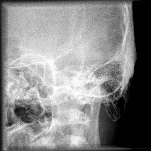
Temporal bone (Stenvers view)
Citation, DOI, disclosures and article data
At the time the article was created Frank Gaillard had no recorded disclosures.
View Frank Gaillard's current disclosuresAt the time the article was last revised Raymond Chieng had no financial relationships to ineligible companies to disclose.
View Raymond Chieng's current disclosures- Stenvers projection
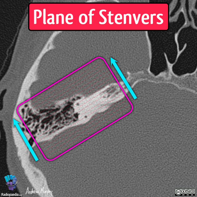
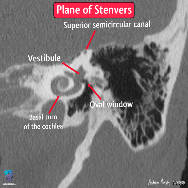
Stenvers view is an oblique radiographic projection used to demonstrate the petrous temporal bone, internal acoustic meatus, and bony labyrinth. Stenvers view is an oblique coronal reconstruction parallel to the petrous portion of the temporal bone. Fine slice multi-detector CT of the petrous bone has replaced the Stenver view due to far superior anatomic detail. It was also used to assess electrode placement following the insertion of a cochlear implant, but has been succeeded by the modified Stenvers view.
The central beam is positioned 45 degrees with the plane of IR and 12 degrees cephalad.
A similar projection can be obtained on reconstructed CT images, approximated by an oblique coronal projection.
History and etymology
The view is named after Hendrik Willem Stenvers (1889–1973), a physician from Utrecht, the Netherlands, who trained in psychiatry and neurology and ran the University Clinic's Röntgen department 2. He developed the now eponymous projection to study pontine angle tumors during the 1910s.
ADVERTISEMENT: Supporters see fewer/no ads
See also
References
- 1. Vogl TJ. Differential Diagnosis in Head and Neck Imaging. Thieme. (1999) ISBN:0865778116. Read it at Google Books - Find it at Amazon
- 2. J A M Frederiks, G W Bruyn and P Eling (Eds.). History of Neurology in the Netherlands. (2002) ISBN: 9789053526866 - Google Books
Incoming Links
Related articles: Radiographs (adult)
- general radiography (adult)
- radiographic positioning terminology[+][+]
-
systematic radiographic technical evaluation (mnemonic)
- portable radiography
- chest radiography[+][+]
- abdominal radiography[+][+]
-
upper limb radiography[+][+]
-
shoulder girdle radiography
- scapula series
-
shoulder series
- shoulder (AP view)
- shoulder (internal rotation view)
- shoulder (external rotation view)
- shoulder (superior-inferior axial view)
- shoulder (inferior-superior axial)
- shoulder (West Point view)
- shoulder (Velpeau view)
- shoulder (modified trauma axial view)
- shoulder (supine lateral view)
- shoulder (modified transthoracic supine lateral)
- shoulder (lateral scapula view)
- shoulder (AP glenoid view)
- shoulder (Garth view)
- shoulder (outlet view)
- shoulder (Stryker notch view)
- acromioclavicular joint series
-
clavicle series
- clavicle (AP view)
- clavicle (AP cephalic view)
- clavicle (oblique view)
- sternoclavicular joint series
- arm and forearm radiography
- wrist and hand radiography
- wrist series
- scaphoid series
- hand series
- thumb series
- fingers series
- rheumatology hands series
- bone age (radiograph)
-
shoulder girdle radiography
-
lower limb radiography[+][+]
- pelvic girdle radiography
- thigh and leg radiography
- ankle and foot radiography
- skull radiography[+][+]
-
paranasal sinus and facial bone radiography
- facial bones[+][+]
- mandible[+][+]
- nasal bone[+][+]
- zygomatic arches[+][+]
- paranasal sinuses[+][+]
- temporal bones
- axiolateral oblique view
- AP axial view
- temporal bone (Stenvers view)
- temporal bone (modified Stenvers view)
- dental radiography[+][+]
- orthopantomography
- temporomandibular joints
- temporomandibular joint (AP axial view)
- temporomandibular joint (axiolateral oblique view)
-
spinal radiography[+][+]
- cervical spine series
-
thoracic spine series
- thoracic spine (AP view)
- thoracic spine (lateral view)
- thoracic spine (oblique view)
- lumbar spine series
- sacrococcygeal radiography


 Unable to process the form. Check for errors and try again.