
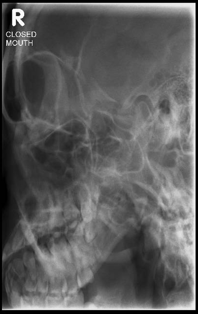
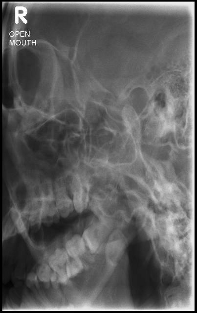
The axiolateral oblique temporomandibular joint (TMJ) view allows for visualization of the articular tubercle, mandibular condyle and fossa of the temporomandibular joint (TMJ).
On this page:
Indications
This projection is useful in identifying structural changes and displaced fractures, assessing excursion and joint spaces in the trauma setting, and evaluating the presence of joint noises, trismus and occlusal alterations 1.
Patient position
the patient is seated upright with the side of interest closest to the detector.
-
the head is placed in a true lateral position
interpupillary line (IPL) perpendicular and midsagittal plane (MSP) parallel to the detector
oblique the body to assist in patient positioning and reduce the object-to-image receptor distance
depending on the projection (open or closed mouth) instruct the patient to open their mouth side and keep it there or keep it shut
Technical factors
left and right lateral and open and closed mouth
-
centering point
central ray 25-30º caudad, centered 5 cm superior and 1 cm anterior to the external auditory meatus
-
collimation
no more than 10 x 10 cm with temporomandibular joint of interest in the middle of the image
-
orientation
portrait
-
detector size
18 cm x 24 cm
-
exposure
70-75 kVp
16-25 mAs
-
SID
100 cm
-
grid
yes
Image technical evaluation
the temporomandibular joint closest to the image receptor should be clearly demonstrated without the superimposition of the opposite temporomandibular joint.
the joint is central on the radiograph
Practical points
a radiolucent support such as a sponge can be used to help maintain the head position
in patients that cannot stand unsupported, this projection can be performed prone to increase patient stability


 Unable to process the form. Check for errors and try again.