
The thoracic spine series is comprised of two standard projections along with a range of additional projections depending on clinical indications. The series is often utilized in the context of trauma, postoperative imaging and for chronic conditions.
Radiographs of the thoracic spine are considered the basic primary imaging, having a far inferior diagnostic yield than that of CT and MRI 1.
Indications
Thoracic spine radiographs are performed for a variety of indications including 1,2:
fall from a height of greater than 3 meters
ejection from a motor vehicle or motorcycle
neurological deficit
postoperative imaging
chronic conditions
history of cancer and associated back pain
Projections
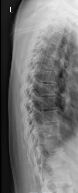
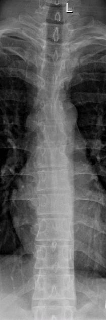
Standard projections
-
images the entirety of the thoracic spine, which consists of twelve vertebrae
intervertebral joints are seen in profile
often performed erect unless otherwise indicated
-
intervertebral joints and neural foramen are open, with the superimposition of the posterior spinous processes
ideal projection when examining for suspected fractures and dislocations
Modified trauma projections
-
horizontal beam lateral
visualization of thoracic vertebral bodies, pedicles, and facet joints taken supine
used in the context of trauma
Additional projections
-
flexion-extension view
functional view used to assess spinal stability
-
bolster view
specialized view for scoliosis, often performed under the guidance of an orthopedic surgeon


 Unable to process the form. Check for errors and try again.