
Segond fracture
Citation, DOI, disclosures and article data
At the time the article was created Frank Gaillard had no recorded disclosures.
View Frank Gaillard's current disclosuresAt the time the article was last revised Lam Van Le had no financial relationships to ineligible companies to disclose.
View Lam Van Le's current disclosures- Segond's fracture
- Lateral capsular sign
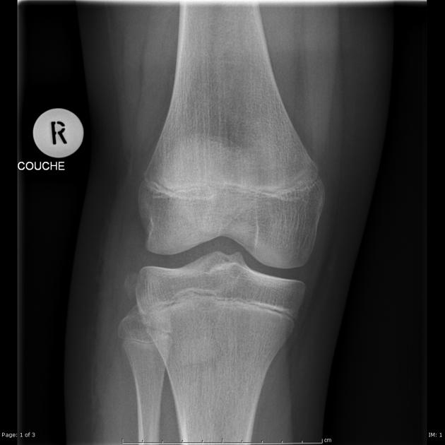
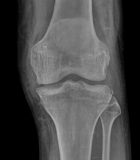
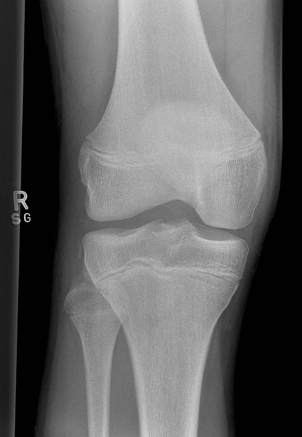
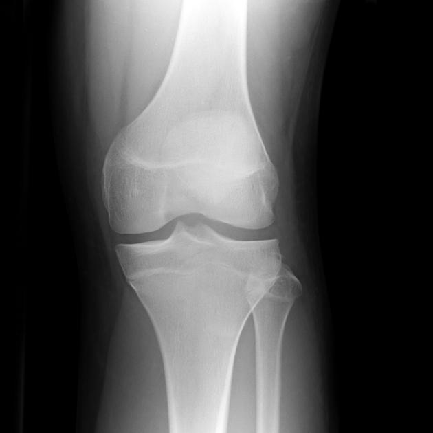
Segond fracture is an avulsion fracture of the knee that involves the lateral aspect of the tibial plateau and is very frequently (~75% of cases) associated with disruption of the anterior cruciate ligament (ACL). On the frontal knee radiograph, it may be referred to as the lateral capsular sign.
On this page:
Clinical presentation
Contrary to the more common causes of an ACL tear, which typically involves valgus stress 3, a Segond fracture usually occurs as a result of internal rotation and varus stress 1,4. Typically these injuries are seen in two settings:
falls
sports: especially soccer, skiing, basketball and baseball 4,10
Pathology
Somewhat surprisingly, the exact cause of a Segond fracture continues to be contentious. The conventional teaching has been that it is the result of avulsion of the middle third of the lateral capsular ligaments 7. Other candidate structures include the iliotibial band and anterior oblique band of the fibular collateral ligament 3.
In 2017, the Anterolateral Ligament Expert Group released a consensus paper 8 recognizing the presence of the anterolateral ligament (ALL) 9 and noted a constant attachment to the lateral meniscus. However, the ALL is inconsistently identified on MRI 7.
A 2017 MRI study of 36 Segond fractures associated with ACL injury showed that the fracture fragment is almost always attached to the posterior fibers of the ITB and the lateral capsule 12. They were unable to demonstrate attachment to the ALL.
Radiographic features
Plain radiograph
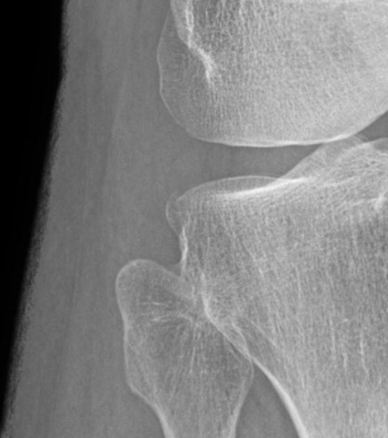
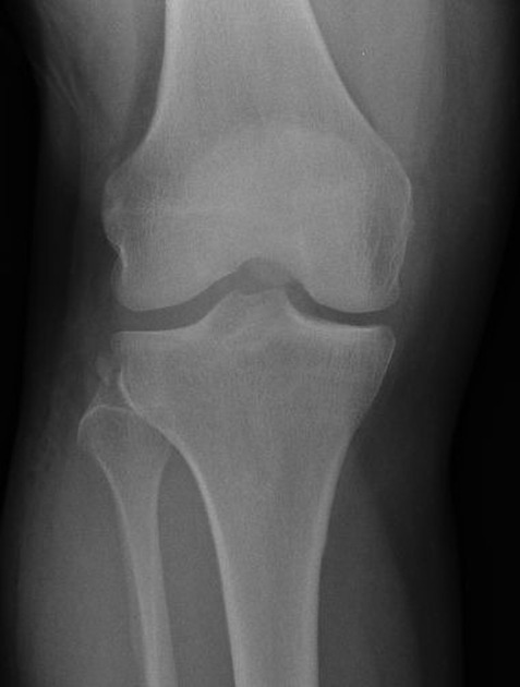
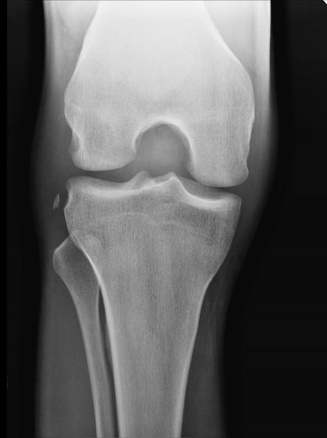
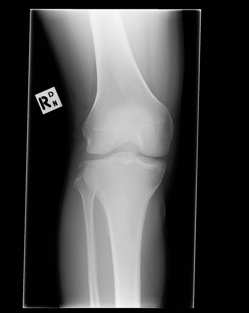
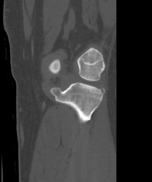
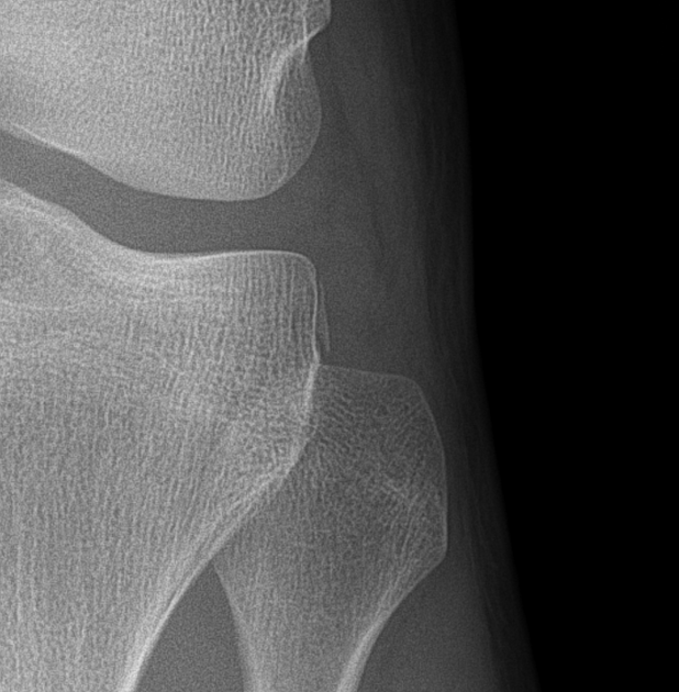
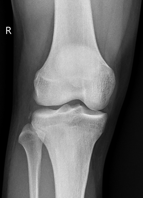
The classical appearance of a Segond fracture is that of a curvilinear or elliptic bone fragment projected parallel to the lateral aspect of the tibial plateau. This has been referred to as the lateral capsular sign 1, which is best seen on the anteroposterior view of the knee.
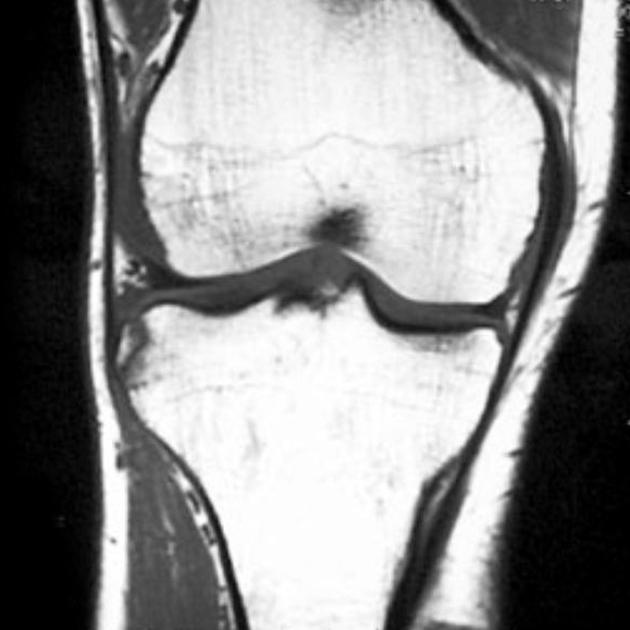
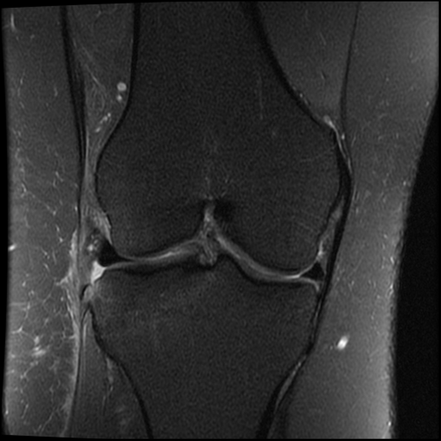
MRI
MRI is essential in all cases of Segond fractures to identify internal derangement. Disruption of the ACL is the most common, however, there are additional frequently encountered injuries. Associated injuries include 1,3:
-
ACL tear
most common associated injury
75-100% of cases 6
-
medial or lateral meniscal tear
66-75% of cases 6
posterior horn most common
avulsion of ACL from the tibial attachment: rare
avulsion of fibular attachment of the long head of biceps femoris
avulsion of the fibular collateral ligament
Treatment and prognosis
Although the fracture itself is small, the extensive ligamentous injury associated with it usually requires surgical intervention, to correct anterior rotational instability 4. Spontaneous healing of the Segond fracture may occur following ACL reconstruction 11 and is associated with a particular bone excrescence arising below the lateral tibial plateau.
History and etymology
First described by Paul Ferdinand Segond, French surgeon (1851-1912) based on cadaveric experiments 1,2,4.
Differential diagnosis
Imaging differential considerations include:
-
arcuate sign: avulsion fracture of the head of the fibula 5
fragment oriented more horizontally
References
- 1. Gottsegen CJ, Eyer BA, White EA et-al. Avulsion fractures of the knee: imaging findings and clinical significance. Radiographics. 2008;28 (6): 1755-70. doi:10.1148/rg.286085503 - Pubmed citation
- 2. Somford MP, Nieuwe Weme RA, Hoornenborg D, Wiegerinck JI, van Raay JJAM, Brouwer RW, Williams A. Biographical background and origin of common eponymous terms in orthopedic surgery: anatomy and fractures in knee surgery. (2018) European journal of orthopaedic surgery & traumatology : orthopedie traumatologie. 28 (1): 79-84. doi:10.1007/s00590-017-2005-x - Pubmed
- 3. Roberts CC, Towers JD, Spangehl MJ et-al. Advanced MR imaging of the cruciate ligaments. Radiol. Clin. North Am. 2007;45 (6): 1003-16, vi-vii. doi:10.1016/j.rcl.2007.08.007 - Pubmed citation
- 4. Goldman AB, Pavlov H, Rubenstein D. The Segond fracture of the proximal tibia: a small avulsion that reflects major ligamentous damage. AJR Am J Roentgenol. 1988;151 (6): 1163-7. AJR Am J Roentgenol (abstract) - Pubmed citation
- 5. Huang GS, Yu JS, Munshi M et-al. Avulsion fracture of the head of the fibula (the "arcuate" sign): MR imaging findings predictive of injuries to the posterolateral ligaments and posterior cruciate ligament. AJR Am J Roentgenol. 2003;180 (2): 381-7. AJR Am J Roentgenol (full text) - Pubmed citation
- 6. Campos JC, Chung CB, Lektrakul N et-al. Pathogenesis of the Segond fracture: anatomic and MR imaging evidence of an iliotibial tract or anterior oblique band avulsion. Radiology. 2001;219 (2): 381-6. Radiology (full text) - Pubmed citation
- 7. Humza Shaikh, Elmar Herbst, Ata Amir Rahnemai-Azar, Marcio Bottene Villa Albers, Jan-Hendrik Naendrup, Volker Musahl, James J. Irrgang, Freddie H. Fu. The Segond Fracture Is an Avulsion of the Anterolateral Complex:. (2017) The American Journal of Sports Medicine. 45 (10): 2247-2252. doi:10.1177/0363546517704845 - Pubmed
- 8. Bertrand Sonnery-Cottet, Matthew Daggett, Jean-Marie Fayard, Andrea Ferretti, Camilo Partezani Helito, Martin Lind, Edoardo Monaco, Vitor Barion Castro de Pádua, Mathieu Thaunat, Adrian Wilson, Stefano Zaffagnini, Jacco Zijl, Steven Claes. Anterolateral Ligament Expert Group consensus paper on the management of internal rotation and instability of the anterior cruciate ligament - deficient knee. (2017) Journal of Orthopaedics and Traumatology. 18 (2): 91. doi:10.1007/s10195-017-0449-8 - Pubmed
- 9. Steven Claes, Evie Vereecke, Michael Maes, Jan Victor, Peter Verdonk, Johan Bellemans. Anatomy of the anterolateral ligament of the knee. (2013) Journal of Anatomy. 223 (4): 321. doi:10.1111/joa.12087 - Pubmed
- 10. Slagstad I, Slagstad PA, Slagstad ST, Slagstad IE, Slagstad. Incidence and Prognostic Significance of the Segond Fracture in Patients Undergoing Anterior Cruciate Ligament Reconstruction. (2020) The American journal of sports medicine. doi:10.1177/0363546520905557 - Pubmed
- 11. Kumahara R, Kimura Y, Sasaki S et al. Prevalence of Segond Fractures Associated with Anterior Cruciate Ligament Injuries and Their Influence on Knee Joint Stability; A Case-Control Study. BMC Musculoskelet Disord. 2022;23(1):180. doi:10.1186/s12891-022-05127-w - Pubmed
- 12. Shaikh H, Herbst E, Rahnemai-Azar A et al. The Segond Fracture Is an Avulsion of the Anterolateral Complex. Am J Sports Med. 2017;45(10):2247-52. doi:10.1177/0363546517704845 - Pubmed
Incoming Links
- Knee radiograph (checklist)
- Anterior cruciate ligament tear
- Eponymous fractures
- Sliver sign (patella)
- Posterolateral corner injury of the knee
- Paediatric knee radiograph (an approach)
- Anterolateral ligament of the knee
- Knee joint
- Reverse Segond fracture
- Avulsion fractures of the knee
- Tibiofemoral joint
- Lateral collateral ligament injury of the knee
- Paediatric curriculum
- Knee radiograph (an approach)
- Arcuate sign (knee)
- Lower extremity fractures
- Distal biceps femoris tendon
- MRI of the knee (an approach)
- Segond fracture
- Complete anterior cruciate ligament tear
- Segond fracture
- Segond fracture
- Segond fracture
- Segond fracture
- Segond fracture
- Avulsion fractures of the knee
- Segond and tibial spine avulsion fractures
- Segond fracture
- Segond fracture
- Segond fracture
- ACL avulsion and Segond fracture
- Segond fracture
- Segond fracture
- Segond fracture
- Segond fracture
- Segond and reverse Segond fractures
- Segond fracture associated with ACL full thickness tear
- Segond fracture with associated anterior cruciate ligament tear
Related articles: Fractures
-
fracture
- terminology[+][+]
- fracture location
- diaphyseal fracture
- metaphyseal fracture
- physeal fracture
- epiphyseal fracture
- fracture types
- avulsion fracture
- articular surface injuries
- complete fracture
- incomplete fracture
- infraction
- compound fracture
- pathological fracture
- stress fracture
- fracture displacement
- fracture location
- fracture healing[+][+]
- skull fractures[+][+]
-
facial fractures[+][+]
- fractures involving a single facial buttress
- alveolar process fractures
- frontal sinus fracture
- isolated zygomatic arch fractures
- mandibular fracture
- nasal bone fracture
- orbital blow-out fracture
- paranasal sinus fractures
- complex fractures
- dental fractures
- fractures involving a single facial buttress
-
spinal fractures[+][+]
- classification (AO Spine classification systems)
-
cervical spine fracture classification systems
- AO classification of upper cervical injuries
- AO classification of subaxial injuries
- Anderson and D'Alonzo classification (odontoid fracture)
- Roy-Camille classification (odontoid process fracture)
- Gehweiler classifcation (atlas fractures)
- Levine and Edwards classification (hangman fracture)
- Allen and Ferguson classification (subaxial spine injuries)
- subaxial cervical spine injury classification (SLIC)
- thoracolumbar spinal fracture classification systems
- three column concept of spinal fractures (Denis classification)
- classification of sacral fractures
-
cervical spine fracture classification systems
- spinal fractures by region
- spinal fracture types
- classification (AO Spine classification systems)
- rib fractures[+][+]
- sternal fractures
-
upper limb fractures[+][+]
- classification
- Rockwood classification (acromioclavicular joint injury)
- AO classification (clavicle fracture)
- Neer classification (clavicle fracture)
- Neer classification (proximal humeral fracture)
- AO classification (proximal humeral fracture)
- AO/OTA classification of distal humeral fractures
- Milch classification (lateral humeral condyle fracture)
- Weiss classification (lateral humeral condyle fracture)
- Bado classification of Monteggia fracture-dislocations (radius-ulna)
- Mason classification (radial head fracture)
- Frykman classification (distal radial fracture)
- Mayo classification (scaphoid fracture)
- Hintermann classification (gamekeeper's thumb)
- Eaton classification (volar plate avulsion injury)
- Keifhaber-Stern classification (volar plate avulsion injury)
- upper limb fractures by region
- shoulder
- clavicular fracture
-
scapular fracture
- acromion fracture
- coracoid process fracture
- glenoid fracture
- humeral head fracture
- proximal humeral fracture
- humeral neck fracture
- arm
- elbow
- forearm
- wrist
-
carpal bones
- scaphoid fracture
- lunate fracture
- capitate fracture
- triquetral fracture
- pisiform fracture
- hamate fracture
- trapezoid fracture
- trapezium fracture
- hand
- shoulder
- classification
- lower limb fractures
- classification by region[+][+]
- pelvic fractures
- hip fractures
- Pipkin classification (femoral head fracture)
- Garden classification (hip fracture)
- American Academy of Orthopedic Surgeons classification (periprosthetic hip fracture)
- Cooke and Newman classification (periprosthetic hip fracture)
- Johansson classification (periprosthetic hip fracture)
- Vancouver classification (periprosthetic hip fracture)
- femoral
- knee
- Schatzker classification (tibial plateau fracture)
- AO classification of distal femur fractures
- Meyers and McKeevers classification (anterior cruciate ligament avulsion fracture)
- tibia/fibula
- Watson-Jones classification (tibial tuberosity avulsion fracture)
- ankle
- foot
- Berndt and Harty classification (osteochondral lesions of the talus)
- Sanders CT classification (calcaneal fracture)
- Hawkins classification (talar neck fracture)
- Myerson classification (Lisfranc injury)
- Nunley-Vertullo classification (Lisfranc injury)
- pelvis and lower limb fractures by region
- pelvic fracture[+][+]
- sacral fracture[+][+]
- coccygeal fracture
-
hip[+][+]
- acetabular fracture
- femoral head fracture
-
femoral neck fracture
- subcapital fracture
- transcervical fracture
- basicervical fracture
-
trochanteric fracture
- pertrochanteric fracture
- intertrochanteric fracture
- subtrochanteric fracture
- femur[+][+]
- mid-shaft fracture
- bisphosphonate-related fracture
- distal femoral fracture
- knee
- avulsion fractures
- Segond fracture
- reverse Segond fracture
- anterior cruciate ligament avulsion fracture
- posterior cruciate ligament avulsion fracture
- arcuate complex avulsion fracture (arcuate sign)
- biceps femoris avulsion fracture
- iliotibial band avulsion fracture
- semimembranosus tendon avulsion fracture
- Stieda fracture (MCL avulsion fracture)
- patellar fracture
- tibial plateau fracture
- avulsion fractures
- leg[+][+]
- tibial tuberosity avulsion fracture
- tibial shaft fracture
- fibular shaft fracture
- Maisonneuve fracture
- ankle[+][+]
- foot[+][+]
- tarsal bones
- metatarsal bones
- phalanges
- classification by region[+][+]
- terminology[+][+]
Related articles: Knee pathology
The knee is a complex synovial joint that can be affected by a range of pathologies:
- bone and cartilage
-
knee fractures
- distal femoral condyle fracture
- tibial plateau fracture (classification)
- patella fracture
-
avulsion fractures of the knee
- arcuate complex avulsion fracture (arcuate sign)
- anterior cruciate ligament avulsion fracture
- biceps femoris avulsion fracture
- iliotibial band avulsion fracture
- quadriceps tendon avulsion fracture
- patellar sleeve fracture
- posterior cruciate ligament avulsion fracture
- reverse Segond fracture
- Segond fracture
- semimembranosus tendon avulsion fracture
- Stieda fracture chronic avulsion injuries[+][+]
- dislocation[+][+]
- chondromalacia patellae
- osteoarthritis of the knee[+][+]
- osteochondral[+][+]
- patterns of bone bruise in knee injury[+][+]
-
knee fractures
- ligaments[+][+]
- anterior cruciate ligament tear
- anterior cruciate ligament ganglion cyst
- anterior cruciate ligament mucoid degeneration
- posterior cruciate ligament tear
- medial collateral ligament tear
- lateral collateral ligament tear
- medial patellofemoral ligament tear
- posterolateral corner injury
- posteromedial corner injury
- tendons[+][+]
- meniscal lesions[+][+]
- bursosynovial lesions[+][+]
- fat pad[+][+]
- popliteal fossa[+][+]
- fascia[+][+]
- alignment[+][+]
- knee
- patellofemoral
- gamut[+][+]


 Unable to process the form. Check for errors and try again.