
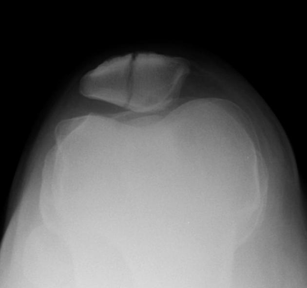
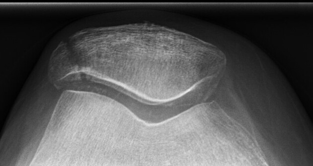
The knee skyline Laurin view is an inferior-superior projection of the patella. It is one of many different methods to obtain an axial projection of the patella.
On this page:
Indication
This view is used in trauma to assess for a patellar fracture or subluxation and in orthopedics for patellofemoral joint disease.1 It is best suited to patients able to maintain a semi-recumbent position on the examination table.
Patient position
the patient is semi-recumbent on the table holding a detector superior of the patella in the landscape orientation
patient's feet should be very close to the tube side of the bed (see technical factors)
the knee is bent close to 30°
often a pillow or cushion should be placed behind the patient to assist them in maintaining this position
Technical factors
inferior-superior axial projection2
-
centering point
the central ray will be angled 160° from the vertical axis (or 30° from horizontal), shooting inferior-superior towards the patella. This will require the tube to lay below the level of the examination table; hence the patient should be as close to this end of the table as possible.
the apex of the patella
-
collimation
laterally to include the skin margins of the knee
inferior to include the femoropatellar joint space
superior to include medial skin margin
-
orientation
landscape
-
detector size
18 cm x 24 cm
-
exposure
60-70 kVp
7-10 mAs
-
SID
100-120 cm
-
grid
no
Image technical evaluation
patella should be free from the superimposition of all bony structures
clear visualization of the patellofemoral joint space
Practical points
This projection is one of the more technically demanding projections of the lower limb. Hence it being one of seven techniques (that the author can find) to achieve it.
This particular method has a high yield if your patients can tolerate the position. Some points to consider when performing this projection:
-
dose
this projection often requires more dose than conventional knee radiographs due to tube angulation and, more often than not, a larger FFD/SID
-
tube angle
30° from horizontal is the academically acceptable angle for this technique, however, assessing the lateral projection and working out the optimal angle from the inferior-superior approach can be beneficial
-
patients feet
the patient's feet will be at the end of the table and often if not careful; the skyline projection may also be a heavily magnified projection of the distal phalanges; ensure the patient's feet are plantar-flexed/out of the primary beam
-
detector
it is possible to use a detector stand rather than asking the patient to hold the detector; this alleviates the risk of motion artifact
-
the pen test
turning the collimator light on, hold the other end of a pen and place it on the lateral border of the patella, if the patient is positioned correctly, the pen will cast a shadow on the detector


 Unable to process the form. Check for errors and try again.