
Primary pulmonary tuberculosis is seen in patients not previously exposed to Mycobacterium tuberculosis.
On this page:
Epidemiology
It is most common in infants and children and has the highest prevalence in children under 5 years of age 1.
Pathology
Infection is the result of droplet inhalation from a close contact which can cause focal pneumonia in a distal airway, typically in the lower lobe. Lymphadenopathy is common. The incubation period can be up to 6 weeks and the infection often remains subclinical, provokes an effective immune response and heals. Occasionally immunity is inadequate and progressive primary TB develops with the potential for miliary spread.
Nucleic acid amplification test and polymerase chain reaction confirm infection and determine rifampin sensitivity. If this is not available, culture, IGRA or tuberculin test conversion can confirm infection.
Clinical Presentation
Fever, loss of weight/failure to thrive, cough or lethargy.
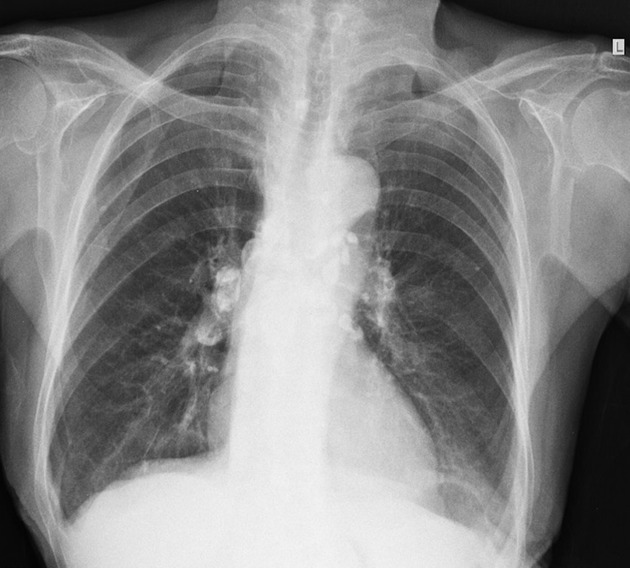
Radiographic features
Primary pulmonary tuberculosis manifests as five main entities:
parenchymal consolidation, often lower zone and typically subpleural 1,2
lymphadenopathy is often the dominant feature and the right hilar and paratracheal nodes are commonly affected 2
clustered parenchymal opacification may give a galaxy sign
miliary opacities indicate progressive disease
Treatment and prognosis
In approximately two-thirds of cases, the parenchymal focus resolves without sequelae at conventional radiography; however, this resolution can take up to 2 years. In the remaining cases, a radiologic scar persists that can calcify in up to 15% of cases, an entity that is known as a Ghon focus. Other calcified foci can also be seen, and persistent mass-like opacities called tuberculomas are seen in ~10% of cases 1.
Complications
progressive primary TB with cavitation
bronchostenosis with atelectasis or overinflation
involvement of the pleura, pericardium or chest wall (empyema necessitans)
involvement of the esophagus or thoracic duct
Differential diagnosis
The appearance of consolidation is indistinguishable from that of bacterial pneumonia.


 Unable to process the form. Check for errors and try again.