5,784 results found
Case
Multifocal hepatocellular carcinoma with portal vein thrombosis

Published
14 Apr 2017
80% complete
CT
Case
Vagal paraganglioma

Published
05 Sep 2021
73% complete
Ultrasound
CT
Case
Rectal cancer - T3

Published
24 Jan 2022
74% complete
MRI
Case
Subependymal giant cell astrocytoma - tuberous sclerosis

Published
25 Jul 2009
77% complete
CT
Case
Cecal and ascending colon cancer

Published
30 Jan 2022
77% complete
CT
Article
Meningeal hemangiopericytoma (historical)
Hemangiopericytomas of the meninges are rare tumors of the meninges, now considered to be an aggressive form of solitary fibrous tumors of the dura. They often present as large and locally aggressive dural masses, frequently extending through the skull vault. They are difficult to distinguish on...
Article
Malignant transformation
Malignant transformation is the term given to the process whereby either normal, metaplastic, or benign neoplastic tissue, becomes a cancer. The process usually occurs in a series of steps and the affected tissue gradually accumulates the genetic mutations that express a malignant phenotype. The...
Case
Large left upper lobe necrotic lung cancer

Published
18 Apr 2011
55% complete
CT
X-ray
Article
Adrenal metastasis
Adrenal metastases are the most common malignant lesions involving the adrenal gland. Metastases are usually bilateral but may also be unilateral. Unilateral involvement is more prevalent on the left side (ratio of 1.5:1).
Epidemiology
They are present at autopsy in up to 27% of patients with ...
Case
Ductal carcinoma in situ recurrence

Published
28 Dec 2013
71% complete
Mammography
Article
Ovarian-Adnexal Reporting and Data System Ultrasound (O-RADS US)
The Ovarian-Adnexal Reporting and Data System Ultrasound (O-RADS US) forms the ultrasound component of the Ovarian-Adnexal Reporting and Data System (O-RADS). This system aims to ensure that there are uniform unambiguous sonographic evaluations of ovarian or other adnexal lesions, accurately ass...
Case
Epithelioid sarcoma - forefoot

Published
01 Dec 2022
77% complete
MRI
Case
Colon cancer (ultrasound)

Published
12 May 2020
81% complete
CT
Ultrasound
Article
Neoplasm
Neoplasms, also known as tumors, are pathological masses, caused by cells abnormally proliferating and/or not appropriately dying. Neoplasms may be either benign or malignant. Malignant neoplasms are synonymous with cancers.
Benign neoplasms
clear origin (unless very large)
slow growth
usua...
Case
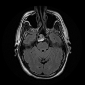
Chordoma - clivus

Published
18 Jan 2022
88% complete
CT
MRI
Article
Basal cell carcinoma
A basal cell carcinoma (BCC) is one of the commonest non-melanocytic types of skin cancer.
Epidemiology
Typically present in elderly fair-skinned patients in the 7th to 8th decades of life. There may be an increased male predilection.
Associations
Multiple basal cell carcinomas may be prese...
Article
Retiform hemangioendothelioma
Retiform hemangioendotheliomas or hobnail hemangioendotheliomas are intermediate locally aggressive and rarely metastasizing vascular neoplasms with a distinctive hobnail endothelial cell morphology.
Epidemiology
Retiform hemangioendotheliomas are rare with <100 cases reported in the literatur...
Case
BI-RADS 5 breast mass - ductal carcinoma

Published
07 Mar 2022
88% complete
Mammography
Case
Metastatic malignant melanoma

Published
28 Nov 2015
92% complete
MRI
CT
Pathology
Article
Kaposiform hemangioendothelioma
Kaposiform hemangioendotheliomas are rare, locally invasive vascular tumors that often present in infancy, most commonly as an enlarging cutaneous mass 1,2. They are currently classified as distinct from tufted angiomas in the ISSVA classification of vascular anomalies. However, some consider it...


 Unable to process the form. Check for errors and try again.