
Blend sign (brain)
Citation, DOI, disclosures and article data
Citation:
Brehmer M, Sharma R, Liu A, et al. Blend sign (brain). Reference article, Radiopaedia.org (Accessed on 13 Mar 2025) https://doi.org/10.53347/rID-42921
rID:
42921
Article created:
15 Feb 2016,
Moritz Brehmer
Disclosures:
At the time the article was created Moritz Brehmer had no recorded disclosures.
View Moritz Brehmer's current disclosures
Last revised:
Disclosures:
At the time the article was last revised Rohit Sharma had no financial relationships to ineligible companies to disclose.
View Rohit Sharma's current disclosures
Revisions:
6 times, by
5 contributors -
see full revision history and disclosures
Systems:
Sections:
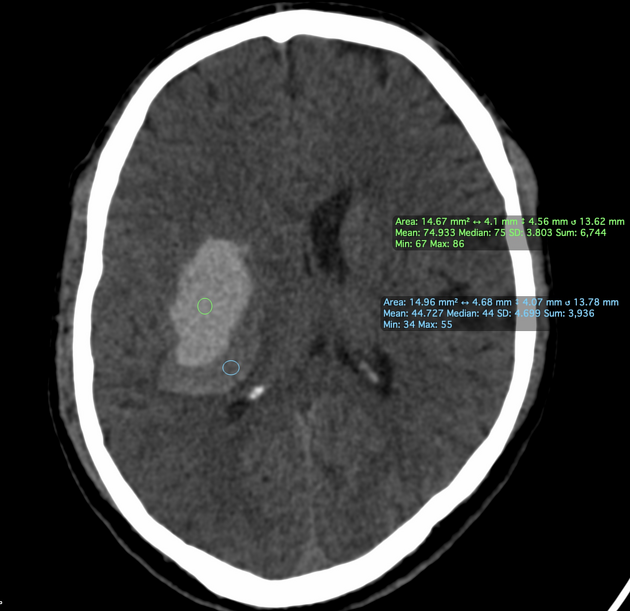
The blend sign refers to the appearance of intracranial hemorrhage in non-contrast CT brain. It is a strong predictor of early hematoma expansion in spontaneous intracerebral hemorrhages, which is a prognosticator for poor functional outcomes 1.
The blend sign is defined as 2:
blending of a relatively hypodense area with an adjacent hyperdense area
a well-defined margin identifiable by the naked eye
at least 18 HU difference between the two areas
the relatively hypodense area not being encapsulated by the hyperdense region
References
- 1. Zhang M, Chen J, Zhan C et al. Blend Sign Is a Strong Predictor of the Extent of Early Hematoma Expansion in Spontaneous Intracerebral Hemorrhage. Front Neurol. 2020;11:334. doi:10.3389/fneur.2020.00334 - Pubmed
- 2. Li Q, Zhang G, Huang Y et al. Blend Sign on Computed Tomography. Stroke. 2015;46(8):2119-23. doi:10.1161/strokeaha.115.009185
Incoming Links
Related articles: Stroke and intracranial haemorrhage
-
stroke and intracranial hemorrhage
- general articles
-
ischemic stroke
- general discussions
- scoring and classification systems
- Alberta stroke program early CT score (ASPECTS)
- ASCOD classification
- Canadian Neurological Scale
- Heidelberg bleeding classification
- NIH Stroke Scale
- Mathew stroke scale
- modified Rankin scale
- Orgogozo Stroke Scale
- Scandinavian Stroke Scale
- thrombolysis in cerebral infarction (TICI) scale
- TOAST classification
- collateral vessel scores
- signs
- by region
- hemispheric infarcts
- frontal lobe infarct
- parietal lobe infarct
- temporal lobe infarct
- occipital lobe infarct
- alexia without agraphia syndrome: PCA
- cortical blindness syndrome (Anton syndrome): top of basilar or bilateral PCA
- Balint syndrome: bilateral PCA
- lacunar infarct
-
thalamic infarct
- artery of Percheron infarct
- Déjerine-Roussy syndrome (thalamic pain syndrome): thalamoperforators of PCA
- top of the basilar syndrome
- striatocapsular infarct
- choroid plexus infarct
- cerebellar infarct
-
brainstem infarct
- midbrain infarct
- Benedikt syndrome: PCA
- Claude syndrome: PCA
- Nothnagel syndrome: PCA
- Weber syndrome: PCA
- Wernekink commissure syndrome
- pontine infarct
- Brissaud-Sicard syndrome
- facial colliculus syndrome
- Gasperini syndrome: basilar artery or AICA
- inferior medial pontine syndrome (Foville syndrome): basilar artery
- lateral pontine syndrome (Marie-Foix syndrome): basilar artery or AICA
- locked-in syndrome: basilar artery
- Millard-Gubler syndrome: basilar artery
- Raymond syndrome: basilar artery
- medullary infarct
- Babinski-Nageotte syndrome
- Cestan-Chenais syndrome
- hemimedullary syndrome (Reinhold syndrome)
- lateral medullary stroke syndrome (Wallenberg syndrome)
- medial medullary syndrome (Déjerine syndrome)
- Opalski syndrome
- midbrain infarct
- acute spinal cord ischemia syndrome
- hemispheric infarcts
- by vascular territory
- by vessel size
- treatment options
- complications
-
intracranial hemorrhage
-
intra-axial hemorrhage
- signs and formulas
- ABC/2 (volume estimation)
- black hole sign
- blend sign
- cashew nut sign
- CTA spot sign
- island sign
- satellite sign
- swirl sign
- zebra sign
- by type
- by location
- signs and formulas
- extra-axial hemorrhage
- extradural hemorrhage (EDH)
- intralaminar dural hemorrhage
- subdural hemorrhage (SDH)
-
subarachnoid hemorrhage (SAH)
- types
- complications
- grading systems
- subpial hemorrhage
-
intra-axial hemorrhage


 Unable to process the form. Check for errors and try again.