Medial medullary syndrome
Citation, DOI, disclosures and article data
At the time the article was created Ahmed Abdrabou had no recorded disclosures.
View Ahmed Abdrabou's current disclosuresAt the time the article was last revised Rohit Sharma had no financial relationships to ineligible companies to disclose.
View Rohit Sharma's current disclosures- Dejerine syndrome
- Dejerine's syndrome
- Déjerine syndrome
- Déjerine's syndrome
Medial medullary syndrome, also known as Déjerine syndrome, is secondary to thrombotic or embolic occlusion of small perforating branches from vertebral or proximal basilar artery supplying the medial aspect of medulla oblongata1,2.
On this page:
Epidemiology
Represents less than 1% of brainstem stroke syndromes 1,2.
Clinical presentation
It is characterized by contralateral hemiplegia/hemiparesis as well as hemisensory loss with ipsilateral hypoglossal palsy (ipsilateral tongue weakness and atrophy) from involvement of CN XII nucleus 1,2. Other manifestations such as vertigo, nausea, or contralateral limb ataxia are also reported 1,2.
Radiographic features
MRI
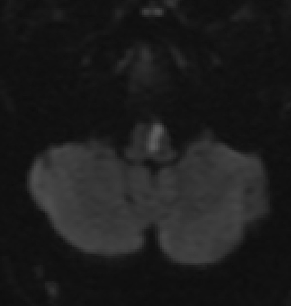
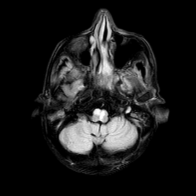
MRI with DWI is the best diagnostic test to confirm the infarct in the medial medulla, whereby the infarcted area has high DWI signal and is low signal on ADC. If bilateral medial medullary infarcts are present, the heart sign may be observed 4.
ADVERTISEMENT: Supporters see fewer/no ads
History and etymology
The syndrome was first described by Joseph Jules Déjerine (1849-1917), a French neurologist, in 1915 3.
References
- 1. Bassetti C, Bogousslavsky J, Mattle H et-al. Medial medullary stroke: report of seven patients and review of the literature. Neurology. 1997;48 (4): 882-90. Pubmed citation
- 2. Ropper AH, Samuels MA, Klein JP. Adams and Victor's principles of neurology 10th ed. New York: McGraw-Hill Medical Pub. Division; 2014.
- 3. Dejerine J. Semiologie des affections du systeme nerveux. The Journal of Nervous and Mental Disease. 1915 Nov 1;42(11):780.
- 4. Duarte-Celada W, Montalvan V, Bueso T, Davila-Siliezar P. Bilateral Medial Medullary Stroke: “The Heart Sign”. Radiology Case Reports. 2024;19(4):1329-32. doi:10.1016/j.radcr.2024.01.008 - Pubmed
Incoming Links
Related articles: Stroke and intracranial haemorrhage
-
stroke and intracranial hemorrhage
- general articles[+][+]
-
ischemic stroke
- general discussions[+][+]
- scoring and classification systems[+][+]
- Alberta stroke program early CT score (ASPECTS)
- ASCOD classification
- Canadian Neurological Scale
- Heidelberg bleeding classification
- NIH Stroke Scale
- Mathew stroke scale
- modified Rankin scale
- Orgogozo Stroke Scale
- Scandinavian Stroke Scale
- thrombolysis in cerebral infarction (TICI) scale
- TOAST classification
- collateral vessel scores
- signs[+][+]
- by region
- hemispheric infarcts[+][+]
- frontal lobe infarct
- parietal lobe infarct
- temporal lobe infarct
- occipital lobe infarct
- alexia without agraphia syndrome: PCA
- cortical blindness syndrome (Anton syndrome): top of basilar or bilateral PCA
- Balint syndrome: bilateral PCA
- lacunar infarct[+][+]
-
thalamic infarct[+][+]
- artery of Percheron infarct
- Déjerine-Roussy syndrome (thalamic pain syndrome): thalamoperforators of PCA
- top of the basilar syndrome
- striatocapsular infarct
- choroid plexus infarct
- cerebellar infarct
-
brainstem infarct
- midbrain infarct[+][+]
- Benedikt syndrome: PCA
- Claude syndrome: PCA
- Nothnagel syndrome: PCA
- Weber syndrome: PCA
- Wernekink commissure syndrome
- pontine infarct[+][+]
- Brissaud-Sicard syndrome
- facial colliculus syndrome
- Gasperini syndrome: basilar artery or AICA
- inferior medial pontine syndrome (Foville syndrome): basilar artery
- lateral pontine syndrome (Marie-Foix syndrome): basilar artery or AICA
- locked-in syndrome: basilar artery
- Millard-Gubler syndrome: basilar artery
- Raymond syndrome: basilar artery
- medullary infarct
- Babinski-Nageotte syndrome
- Cestan-Chenais syndrome
- hemimedullary syndrome (Reinhold syndrome)
- lateral medullary stroke syndrome (Wallenberg syndrome)
- medial medullary syndrome (Déjerine syndrome)
- Opalski syndrome
- midbrain infarct[+][+]
- acute spinal cord ischemia syndrome[+][+]
- hemispheric infarcts[+][+]
- by vascular territory[+][+]
- by vessel size[+][+]
- treatment options[+][+]
- complications[+][+]
-
intracranial hemorrhage[+][+]
-
intra-axial hemorrhage
- signs and formulas
- ABC/2 (volume estimation)
- black hole sign
- blend sign
- cashew nut sign
- CTA spot sign
- island sign
- satellite sign
- swirl sign
- zebra sign
- by type
- by location
- signs and formulas
- extra-axial hemorrhage
- extradural hemorrhage (EDH)
- intralaminar dural hemorrhage
- subdural hemorrhage (SDH)
-
subarachnoid hemorrhage (SAH)
- types
- complications
- grading systems
- subpial hemorrhage
-
intra-axial hemorrhage


 Unable to process the form. Check for errors and try again.