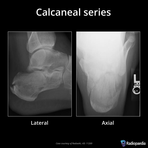
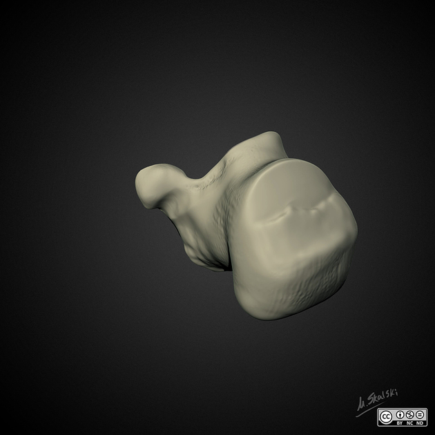
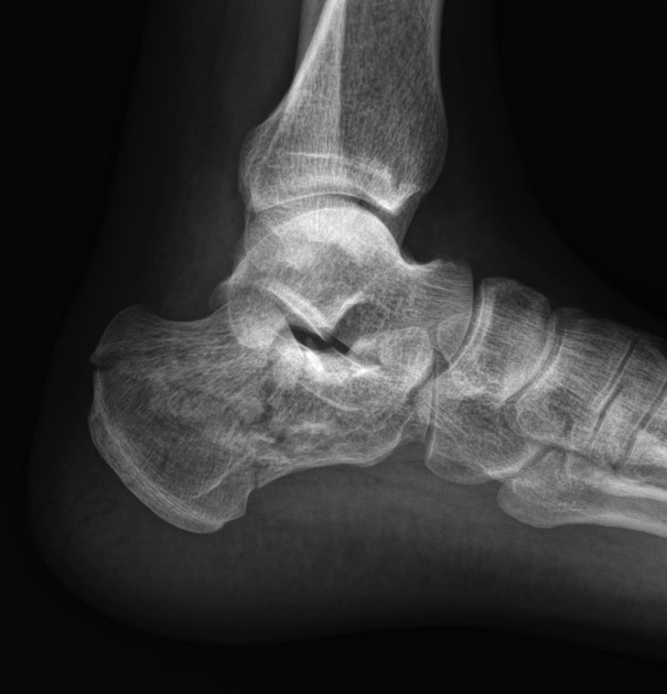
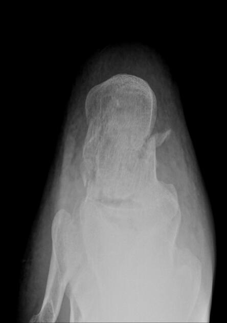
The calcaneus series is comprised of a lateral and axial (plantodorsal) projection. The calcaneus is the most commonly fractured tarsal bone accounting for ~60% of all tarsal fractures 1. This series provides a two view investigation of the calcaneus alongside the talar articulations and talocalcaneal joint.
This series provides the first step in the diagnosis of acute calcaneal fractures. Often the series will be the benchmark in the diagnosis of osseous injuries 3, however, due to the complexity of the region, calcaneus injuries will go on to have further imaging such as computed tomography to define the intricacies of the pathology.
Significant fractures of the calcaneus are resultant of high-energy axial loading; this mechanism has a high association with burst fractures of the lumbar spine 4.
Indications
Calcaneus radiographs are performed for a variety of indications including:
- fall from a significant height onto feet
- axial loading on the talus during deceleration in a motor vehicle accident
- inability to weight bear
- violent twisting injuries
- pathological processes such as osteoporosis, tumors or cysts 2
Projections
Standard projections
- lateral view
-
axial view (Harris)
- the plantar aspect is entirely visual from the posterior tuberosity to the articulation between the calcaneus and talus
Additional projections
- An ankle series and/or foot series may be required after the initial investigation to rule out further pathology.
-
Broden's view
- used to evaluate the posterior subtalar joint
- Canale/Kelly view
- used to evaluate the talar neck
- anterior calcaneal process and calcaneocuboid joint well visualized (also calcaneonavicular coalition)


 Unable to process the form. Check for errors and try again.