
Dysphagia
Updates to Article Attributes
Dysphagia refers to subjective awareness of difficulty or obstruction during swallowing.
Dysphagia is a relatively common and increasingly prevalent clinical problem.
Epidemiology
Dysphagia is common in older age groups2
Women are more prone to have dysphagia than men (80% versus 20%)2
Pathology
Dysphagia may be classified depending on the location of this sensation as oropharyngeal or substernal.
Dysphagia can be caused by functional or structuralabnormalities of the oral cavity, pharynx, esophagus, and/or gastric cardia.
Imaging
Fluoroscopic studies
- modified barium swallow , used for the evaluation of swallowing mechanisms specifically for aspiration or penetration, this exam is usually performed in conjunction with a swallowing therapist to assess swallowing function and response to therapeutic strategies 4
- barium swallow /esophagography provides anatomic and functional information about the pharynx, esophagus, gastroesophageal junction (GEJ), and gastric cardia, including evaluation of esophageal motility and assessment for gastroesophageal reflux
- barium tablet may be used to detect subtle areas of esophageal narrowing 5. The tablet (of known 12.5mm diameter) should be swallowed with a small amount of water and passage is observed at fluoroscopy. If the tablet becomes lodged in a particular location the patient should swallow a small amount of additional water. If the tablet remains lodged, a more detailed assessment should be performed
Manometry can help evaluate the esophageal motor pattern and lower esophageal sphincter function.1
Cross-sectional imaging may be used especially If there's a mass effect on the esophagus seen at esophagography or for evaluation of esophageal tumors.
Classification
Oropharyngeal dysphagia
orophatyngeal dysphagia occurs when a patient symptomatically localizes a sensation of difficulty passing foodblockage in the throat, It can be caused by:
Functional causes
-
laryngeal penetration (when contrast seen entering the larynx at fluoroscopy) or
liquid from the mouthaspiration(when contrast extends inferiorly through thepharynxtrue vocal), which are common with patients who have a history of neurologic disorders including stroke - cricopharyngeal muscle spasm
Structural causes:
- oesophageal web,
- oesophageal diverticula, e.g. Zenker, Killian-Jamieson, and epiphrenic diverticula
- tumors,
- extrinsic masses e.g. thyroid disease, or cervical spine disease.
Substernal dysphagia
substernal dysphagia occurs when a patient symptomatically localizes a sensation of discomfort or blockage between the thoracic inlet and into the stomach
Causes of dysphagia It can be divided broadly into mechanicalcaused by:
Functional causes
-
diffuse oesophageal spasm (DES), characterized by multiple spontaneous and
neuromuscular (motor).uncoordinated esophageal contractions which have the classic "Corkscrew" appearance at esophagogram - achalasia, characterized by esophageal dilatation with distal tapered beaklike narrowing
- scleroderma, characterized by esophageal dilatation with a patulous gastroesophageal junction
Structural causes
See also
- Oesophageal stricture:
- Peptic strictures most often typically appear as smooth, tapered narrowing in the distal esophagus
- Barrett oesophagus occurs often as a consequence of GERD in the mid- to upper esophagus,
- ring stricture, Schatzki ring is the most common type of esophageal ring, associated with a hiatal hernia
- other less common causes of benign strictures include corrosive oesophagitis,Crohn disease, Behçet disease, and eosinophilic esophagitis
- extrinsic mass such as lung cancer, or vascular compression e.g. aberrant right subclavian artery (e.g. dysphagia lusoria)
- esophageal Infections,such as candidiasis, herpes virus, human immunodeficiency virus (HIV), and cytomegalo virus (CMV)
-
oesophageal
stricturecarcinoma can appear infiltrative, ulcerative, polypoid, and/or varicoid at esophagography - impacted foreign body or food bolus in the esophagus. Water-soluble contrast should be used for increased risk of perforation
-<p><strong>Dysphagia</strong> refers to the sensation of difficulty passing food or liquid from the mouth through the pharynx and <a href="/articles/oesophagus_(textbook)">oesophagus</a> and into the <a href="/articles/stomach">stomach</a>. </p><p>Causes of dysphagia can be divided broadly into mechanical and neuromuscular (motor).</p><h4>See also</h4><ul>-<li><a href="/articles/achalasia">achalasia</a></li>-<li><a href="/articles/presbyoesophagus">presbyoesophagus</a></li>-<li><a href="/articles/oesophageal_web">oesophageal web</a></li>-<li><a href="/articles/oesophageal_cancer">oesophageal cancer</a></li>-<li><a href="/articles/oesophageal_stricture">oesophageal stricture</a></li>-</ul>- +<p><strong>Dysphagia</strong> refers to subjective awareness of difficulty or obstruction during swallowing.</p><p>Dysphagia is a relatively common and increasingly prevalent clinical problem.</p><h4>Epidemiology</h4><p>Dysphagia is common in older age groups<sup>2</sup></p><p>Women are more prone to have dysphagia than men (80% versus 20%)<sup>2</sup></p><h4>Pathology</h4><p>Dysphagia may be classified depending on the location of this sensation as oropharyngeal or substernal.</p><p>Dysphagia can be caused by functional or structural<em> </em>abnormalities of the oral cavity, pharynx, esophagus, and/or gastric cardia.</p><h4>Imaging</h4><p><strong>Fluoroscopic studies</strong></p><ul>
- +<li>
- +<em>modified barium swallow</em> , used for the evaluation of swallowing mechanisms specifically for aspiration or penetration, this exam is usually performed in conjunction with a swallowing therapist to assess swallowing function and response to therapeutic strategies <sup>4</sup>
- +</li>
- +<li>
- +<em><a href="/articles/barium-swallow">barium swallow</a> /esophagography</em> provides anatomic and functional information about the pharynx, esophagus, gastroesophageal junction (GEJ), and gastric cardia, including evaluation of esophageal motility and assessment for gastroesophageal reflux</li>
- +<li>
- +<em>barium tablet</em> may be used to detect subtle areas of esophageal narrowing <sup>5</sup>. The tablet (of known 12.5mm diameter) should be swallowed with a small amount of water and passage is observed at fluoroscopy. If the tablet becomes lodged in a particular location the patient should swallow a small amount of additional water. If the tablet remains lodged, a more detailed assessment should be performed</li>
- +</ul><p><strong>Manometry </strong>can help evaluate the esophageal motor pattern and lower esophageal sphincter function.<sup>1</sup></p><p><strong>Cross-sectional </strong>imaging may be used especially If there's a mass effect on the esophagus seen at esophagography or for evaluation of esophageal tumors.</p><h4>Classification</h4><h5>Oropharyngeal dysphagia</h5><p>orophatyngeal dysphagia occurs when a patient symptomatically localizes a sensation of blockage in the throat, It can be caused by:</p><p><strong>Functional causes</strong></p><ul>
- +<li>laryngeal penetration (when contrast seen entering the larynx at fluoroscopy) or aspiration(when contrast extends inferiorly through the true vocal), which are common with patients who have a history of neurologic disorders including stroke</li>
- +<li><a href="/articles/cricopharyneal-muscle-spasm">cricopharyngeal muscle spasm</a></li>
- +</ul><p><strong>Structural causes:</strong></p><ul>
- +<li>
- +<a href="/articles/oesophageal-web">oesophageal web</a>,</li>
- +<li>
- +<a href="/articles/oesophageal-diverticula">oesophageal diverticula</a>, e.g. <a href="/articles/zenker-diverticulum">Zenker</a>, Killian-Jamieson, and <a href="/articles/epiphrenic-diverticulum">epiphrenic</a> diverticula</li>
- +<li>tumors,</li>
- +<li>extrinsic masses e.g. thyroid disease, or cervical spine disease.</li>
- +</ul><h5>Substernal dysphagia</h5><p>substernal dysphagia occurs when a patient symptomatically localizes a sensation of discomfort or blockage between the thoracic inlet and the xiphoid process. It can be caused by:</p><p><strong>Functional causes</strong></p><ul>
- +<li>
- +<a href="/articles/diffuse-oesophageal-spasm">diffuse oesophageal spasm</a> (DES), characterized by multiple spontaneous and uncoordinated esophageal contractions which have the classic "Corkscrew" appearance at esophagogram </li>
- +<li> <a href="/articles/achalasia">achalasia</a>, characterized by esophageal dilatation with distal tapered beaklike narrowing</li>
- +<li> <a href="/articles/scleroderma">scleroderma</a>, characterized by esophageal dilatation with a patulous gastroesophageal junction </li>
- +</ul><p><strong>Structural causes </strong></p><ul>
- +<li> <a href="/articles/oesophageal-stricture">Oesophageal stricture</a>:<ul>
- +<li>Peptic strictures most often typically appear as smooth, tapered narrowing in the distal esophagus</li>
- +<li>
- +<a href="/articles/barrett-oesophagus">Barrett oesophagus</a> occurs often as a consequence of <a href="/articles/gastro-oesophageal-reflux-disease">GERD</a> in the mid- to upper esophagus,</li>
- +<li>ring stricture, <a href="/articles/schatzki-ring">Schatzki ring</a> is the most common type of esophageal ring, associated with a <a href="/articles/hiatus-hernia">hiatal hernia</a>
- +</li>
- +<li>other less common causes of benign strictures include <a href="/articles/corrosive-oesophagitis">corrosive oesophagitis</a>,<a href="/articles/crohn-disease-1">Crohn </a>disease, Behçet disease, and eosinophilic esophagitis</li>
- +</ul>
- +</li>
- +<li>extrinsic mass such as lung cancer, or vascular compression e.g. aberrant right subclavian artery (e.g. <a href="/articles/dysphagia-lusoria">dysphagia lusoria</a>)</li>
- +<li>esophageal Infections,such as candidiasis, herpes virus, human immunodeficiency virus (HIV), and cytomegalo virus (CMV)</li>
- +<li>
- +<a href="/articles/oesophageal-carcinoma-1">oesophageal carcinoma</a> can appear infiltrative, ulcerative, polypoid, and/or varicoid at esophagography</li>
- +<li> impacted foreign body or food bolus in the esophagus. Water-soluble contrast should be used for increased risk of perforation</li>
- +</ul><p> </p>
References changed:
- 4. Domenech E, Kelly J. Swallowing disorders. Med. Clin. North Am. 1999;83 (1): 97-113, ix. <a href="http://www.ncbi.nlm.nih.gov/pubmed/9927963">Pubmed citation</a><span class="auto"></span>
- 5. Luedtke P, Levine MS, Rubesin SE et-al. Radiologic diagnosis of benign esophageal strictures: a pattern approach. Radiographics. 2003;23 (4): 897-909. <a href="http://dx.doi.org/10.1148/rg.234025717">doi:10.1148/rg.234025717</a> - <a href="http://www.ncbi.nlm.nih.gov/pubmed/12853664">Pubmed citation</a><span class="auto"></span>
- 1. Carucci LR, Turner MA. Dysphagia revisited: common and unusual causes. Radiographics.35 (1): 105-22. <a href="http://dx.doi.org/10.1148/rg.351130150">doi:10.1148/rg.351130150</a> - <a href="http://www.ncbi.nlm.nih.gov/pubmed/25590391">Pubmed citation</a><span class="auto"></span>
- 3. Kuo P, Holloway RH, Nguyen NQ. Current and future techniques in the evaluation of dysphagia. J. Gastroenterol. Hepatol. 2012;27 (5): 873-81. <a href="http://dx.doi.org/10.1111/j.1440-1746.2012.07097.x">doi:10.1111/j.1440-1746.2012.07097.x</a> - <a href="http://www.ncbi.nlm.nih.gov/pubmed/22369033">Pubmed citation</a><span class="auto"></span>
- 2. Wilkins T, Gillies RA, Thomas AM et-al. The prevalence of dysphagia in primary care patients: a HamesNet Research Network study. J Am Board Fam Med. 2007;20 (2): 144-50. <a href="http://dx.doi.org/10.3122/jabfm.2007.02.060045">doi:10.3122/jabfm.2007.02.060045</a> - <a href="http://www.ncbi.nlm.nih.gov/pubmed/17341750">Pubmed citation</a><span class="auto"></span>
- 1. Fauci A, Braunwald E, Kasper D et-al. Harrison's manual of medicine. McGraw-Hill Professional. ISBN:0071477438. <a href="http://books.google.com/books?vid=ISBN0071477438">Read it at Google Books</a> - <a href="http://www.amazon.com/gp/product/0071477438">Find it at Amazon</a><span class="auto"></span>
Image ( create )
Image ( create )
Image 1 Fluoroscopy ( create )

Image 2 Fluoroscopy (Lateral) ( create )

Image 3 X-ray (Oblique) ( create )

Image 4 CT (C+ portal venous phase) ( create )

Image 5 Fluoroscopy ( create )
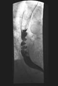
Image 6 Barium ( create )

Image 7 CT (lung window) ( create )

Image 8 Fluoroscopy ( create )

Image 9 Fluoroscopy ( create )
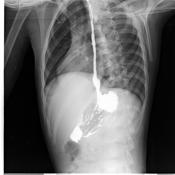
Image 10 CT (C+ arterial phase) ( create )

Image 11 CT (non-contrast) ( create )


 Unable to process the form. Check for errors and try again.