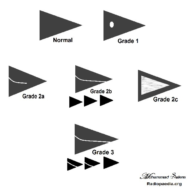
MRI grading system for abnormal meniscal signal intensity
Citation, DOI, disclosures and article data
At the time the article was created Mohammad Salem Amer had no recorded disclosures.
View Mohammad Salem Amer's current disclosuresAt the time the article was last revised Calum Worsley had no financial relationships to ineligible companies to disclose.
View Calum Worsley's current disclosures- MR grading system for meniscal signal intensity
- Grading system for meniscal signal intensity: MRI
- Meniscal signal intensity: MRI grading system
MRI grading system for abnormal high meniscal signal intensity was reported by Lotysch et al.
Classification
Grade 1 to 3 have been described on MRI:
grade 1: small focal area of hyperintensity, no extension to the articular surface
-
grade 2: linear areas of hyperintensity, no definite extension to the articular surface
2a: linear abnormal hyperintensity with no extension to the articular surface
2b: abnormal hyperintensity reaches the articular surface on a single image
2c: globular wedge-shaped abnormal hyperintensity with no extension to the articular surface
grade 3: abnormal hyperintensity extends to at least one articular surface (superior or inferior), on more than one consecutive image, and is referred to as a definite meniscal tear
Grade 2 meniscal signal was found to be associated with a meniscal tear on arthroscopy. Therefore, grade 2 was further subdivided into 2a, 2b, and 2c. Dillon et al. found that 50% of patients with grade 2c had meniscal tears on arthroscopy.
References
- 1. Li C, Kim M, Kim I, Lee J, Jang K, Lee S. Correlation of Histological Examination of Meniscus with MR Images: Focused on High Signal Intensity of the Meniscus Not Caused by Definite Meniscal Tear and Impact on Mr Diagnosis of Tears. Korean J Radiol. 2013;14(6):935-45. doi:10.3348/kjr.2013.14.6.935 - Pubmed
- 2. Lotysch M, Mink J, Crues JV, Schwartz SA. Magnetic resonance imaging in the detection of meniscal injuries (abstract). Magn Reson Imaging 1986;4:185.
- 3. Dillon E, Pope C, Jokl P, Lynch K. The Clinical Significance of Stage 2 Meniscal Abnormalities on Magnetic Resonance Knee Images. Magn Reson Imaging. 1990;8(4):411-5. doi:10.1016/0730-725x(90)90049-8 - Pubmed
Incoming Links
Related articles: Knee pathology
The knee is a complex synovial joint that can be affected by a range of pathologies:
- bone and cartilage
-
knee fractures
- distal femoral condyle fracture
- tibial plateau fracture (classification)
- patella fracture
-
avulsion fractures of the knee
- arcuate complex avulsion fracture (arcuate sign)
- anterior cruciate ligament avulsion fracture
- biceps femoris avulsion fracture
- iliotibial band avulsion fracture
- quadriceps tendon avulsion fracture
- patellar sleeve fracture
- posterior cruciate ligament avulsion fracture
- reverse Segond fracture
- Segond fracture
- semimembranosus tendon avulsion fracture
- Stieda fracture chronic avulsion injuries
- dislocation
- chondromalacia patellae
- osteoarthritis of the knee
- osteochondral
- patterns of bone bruise in knee injury
-
knee fractures
- ligaments
- anterior cruciate ligament tear
- anterior cruciate ligament ganglion cyst
- anterior cruciate ligament mucoid degeneration
- posterior cruciate ligament tear
- medial collateral ligament tear
- lateral collateral ligament tear
- medial patellofemoral ligament tear
- posterolateral corner injury
- posteromedial corner injury
- tendons
- meniscal lesions
- bursosynovial lesions
- fat pad
- popliteal fossa
- fascia
- alignment
- knee
- patellofemoral
- gamut


 Unable to process the form. Check for errors and try again.