
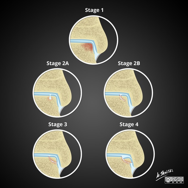
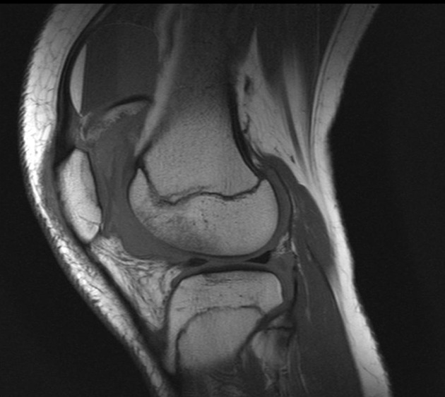
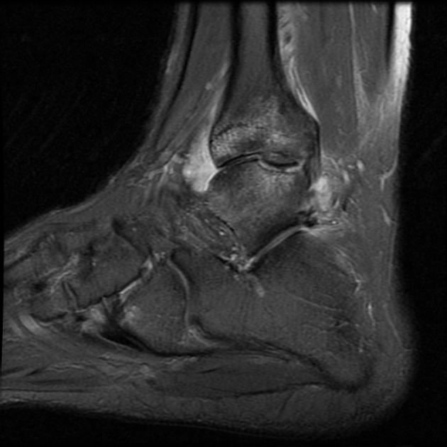
Osteochondral injury staging system for MRI attempts to grade the stability and severity of osteochondral injury and is used to plan management.
Classification
-
stage I
injury limited to articular cartilage
MRI findings: subchondral edema
x-ray findings: none
-
stage II
cartilage injury with associated subchondral fracture but without detachment
thin sclerotic margin
x-ray findings: usually none; may see fracture as sclerotic or osteopenic area
-
two subtypes 2,3
type A: cystic on CT and/or edema on MRI
type B: non-displaced and incompletely undercut by fluid (MR) or lucency (CT), with an open connection to the articular cartilage (essentially 2a without edema on MRI)
-
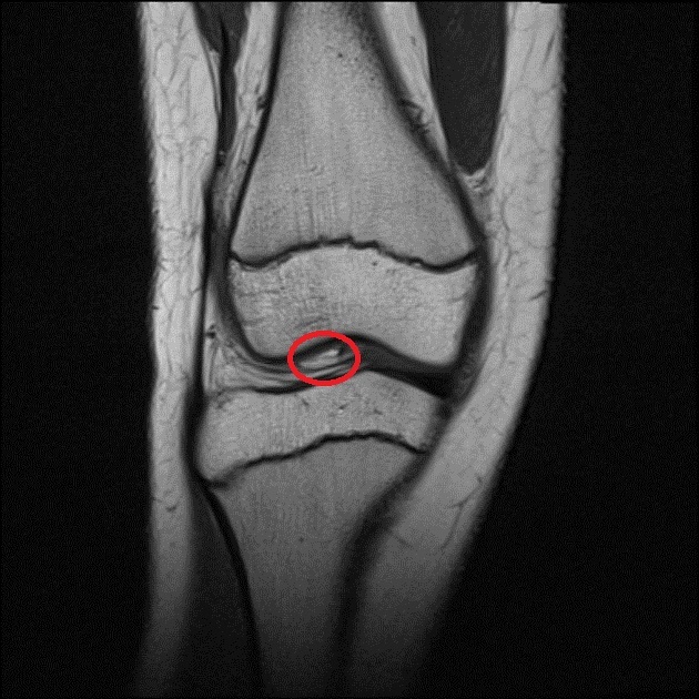
stage III
detached, non-displaced fragment
MRI findings: high signal around osteochondral fracture (rim sign) but not displaced
x-ray findings: slight lucency between osteochondral fragment and remainder of the bone
-
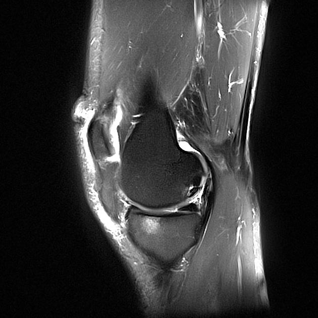
stage IV
osteochondral fragment displaced
usually joint effusion present, surrounding fragment and filling donor site
x-ray findings: increased lucency between osteochondral fragment and remainder of the bone, or loose body with donor site irregularity
-
stage V
subchondral cyst formation
secondary degenerative change
x-ray findings: secondary osteoarthritis


 Unable to process the form. Check for errors and try again.