
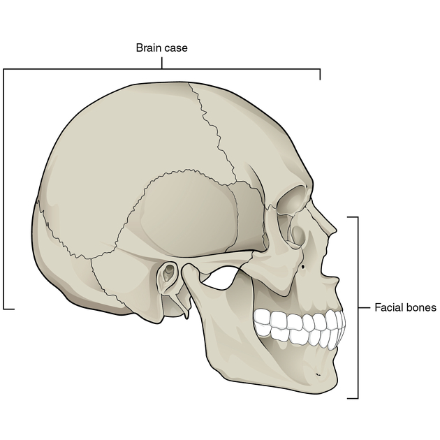
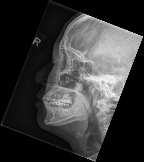
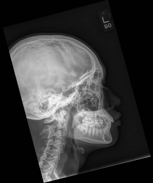
The lateral paranasal sinuses and facial bones view is a nonangled lateral radiograph showcasing the facial bones (i.e. mandible, maxilla, zygoma, nasal, and lacrimal bone) and paranasal sinuses.
On this page:
Indications
This view is useful in assessing any inflammatory processes or fractures to the facial bones, orbits, and paranasal sinuses. In instances of facial trauma, carrying out this view using a horizontal beam can be beneficial in assessing fluid-filled sinus cavities. It is also an important view for localizing foreign bodies in the trauma setting.
Patient position
- rest the lateral aspect of the patient's head (with the side of interest) against the image detector
- adjust the head into a true lateral position, with the midsagittal plane (Figure 1) parallel to the image detector
- for patient's comfort, adjust the patient's body into an oblique position
Technical factors
- lateral projection
-
centering point
- zygoma (cheek prominence), midway between outer canthus and the external auditory meatus (EAM)
-
collimation
- superior to include frontal sinus
- inferior to include chin (especially important to include soft tissue to assess for foreign body or air in a trauma setting)
- anterior to include skin margins
- posterior to the EAM
-
orientation
- landscape
-
detector size
- 24 cm x 30 cm
-
exposure
- 70-80 kVp
- 20-25 mAs
-
SID
- 100 cm
-
grid
- no
Image technical evaluation
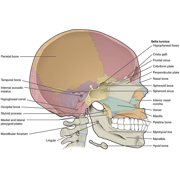
- sella turcica in profile
- temporomandibular joints are superimposed
Practical points
- remove earrings, glasses, piercings, and dentures to avoid artifact obscuring important pathology
- encourage patients to raise their chin to prevent superimposing the mandibular angle over the anterior cervical spine
- to ensure true lateral, the floor of the anterior cranial fossa should be superimposed


 Unable to process the form. Check for errors and try again.