
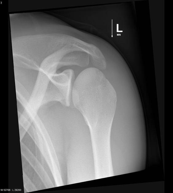
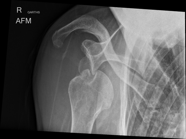
The apical oblique projection or the Garth view of the shoulder is the tangential projection of the shoulder used in trauma 4.
On this page:
Indications
The view is best for evaluating the glenohumeral joint for dislocations and trauma to the glenoid of the scapula; this projection can be used as a replacement to the lateral scapula view in trauma, however, interpretation is difficult. The angle of the beam means it is tangential to the anterior-inferior glenoid rim (great for Bankart fractures) and gives a better view of the posterior humeral head (ideal for Hill-Sachs defect) 1,3.
Patient position
- the patient is preferably erect, best placed on a seat sitting against the upright bucky (due to the angle of the tube)
- the midcoronal plane of the patient is parallel to the image receptor, in other words, the patient's back is against the image receptor
- the glenohumeral joint of the affected side is at the center of the image receptor
- the patient is turned toward the affected side to show the glenohumeral joint space; this is achieved by rotating the patient 30-45°
- if possible the patient has the affected side's hand resting on the unaffected shoulder
Technical factors
- axial oblique projection
-
centering point
- 45° caudal angle of the x-ray tube
- 2.5 cm inferior to the coracoid process, or 2 cm inferior to the lateral clavicle at the level of the glenohumeral joint
-
collimation
- superior to the skin margins
- inferior to include one-third of the proximal humerus
- lateral to include the lateral portion of the humeral head often the skin margins (dependent on body habitus)
- medial to 1/3 of the medial clavicle
-
orientation
- portrait
-
detector size
- 18 cm x 24 cm
-
exposure
- 60-70 kVp
- 10-18 mAs
-
SID
- 100 cm
-
grid
- yes (this can vary departmentally)
Image technical evaluation
- the humeral head will appear elongated (due to angle)
- the coracoid process is sometimes projected over the humeral head
- the AC joint should be superior to the humeral head
-
posterior dislocation
- the humeral head will be projected superior to the glenoid often obstructed by the acromion
-
anterior dislocation
- in the majority of cases, the humeral head will be projected inferior to the glenoid
Practical points
- due to the steep angle of the tube, it is advisable to have the patient sitting on a stool for the examination, otherwise, you may hit the ceiling with your x-ray tube
- use of the AEC is not advisable; best to set your own exposures based on the AP shoulder.
- if possible, angle the detector to prevent elongation


 Unable to process the form. Check for errors and try again.