Radiopaedia Blog

I am constantly concerned by how often the term juxtacortical, in the context of multiple sclerosis, is misunderstood by clinicians, radiologists and radiology residents / registrars. This is particularly concerning as the imaging diagnosis is dependent on understanding this term. I recently added a short article (here) trying to get to the bottom of this definitional issue, but thought I would elaborate a little here. 

The term juxtacortical is particularly important in the context of McDonald criteria, although oddly enough the term is not defined in either the original 2001 paper or the 2010 revision. Perhaps partly due to this, it is a term often misunderstood and thought to mean merely "sort of near the cortex".

Subcortical u-fibers (black) with a juxtacortical lesion (JC) *


This is not the case, but rather it refers to lesions which abut the cortex, and thus involve the subcortical u-fibers (also known as short association fibers and represents connections between adjacent gyri of the brain, located within the cortex or immediately deep to it in the very outer parts of the subcortical white matter). 

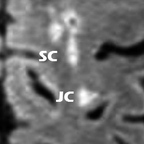
Juxtacortical (JC) vs subcortical (SC) lesions in a patient with MS (whole case here)


An alternative term to juxtacortical which is perhaps more easily understood is leukocortical (i.e. involving both cortex and juxtacortical white matter), especially as the importance of grey matter involvement in multiple sclerosis is growing, and is increasingly imaged using modern sequences (e.g. double inversion recovery) and higher field strength. 

* Image of cortex adapted illustration by Patrick J Lynch. Original wikimediacommons file here.


Dr Frank Gaillard is a neuroradiologist at the Royal Melbourne Hospital, Melbourne, Australia, and is the Founder and Editor of Radiopaedia.org. 

NB: Opinions expressed are those of the author alone, and are not those of his employer, or of Radiopaedia.org

Update

Updated: Dec 29th 2014

We have finished re-processing all images. Phew. That took a while. 


Updated: Dec 5th 2014

Excellent news... we have updated our image processing pipeline and now images you upload will stay just as sharp and awesome as the ones you saw on your PACS. We are also embarking on reprocessing all affected images, so over time all your cases will look as good as they should. Best of all, you don't need to do anything. 


Original post

Original post: Nov 17th 2014

Some of you may have noticed that some of the cases you upload don't look as good as your original version. This is a problem we are aware of and is the inadvertent result of updates we were required to make to improve the speed of serving images - many of your cases have hundreds of images. 


Imaging quality is of course extremely important to us and the current situation is unacceptable. It has taken us some time as many other behind-the-scenes changes have been necessary. The good news is we have been working on it and a fix is almost ready. 

"we will also be going back to all existing affected cases and reprocessing them"

As soon as the fix is deployed (we expect this to be by mid December) all new cases will look awesome. The even better news is that we will also be going back to all existing affected cases and reprocessing them. Thus you can keep uploaded safe in the knowledge that all your existing cases will also look great, even if at the moment they are affected. We can do this because we keep the original images you upload. Of course re-processing literally millions of images will take some time, so please be patient. 

I apologise for this, but at the end of it all you will have great looking cases which load super fast. 

Frank

Dr Frank Gaillard is a neuroradiologist at the Royal Melbourne Hospital, Melbourne, Australia, and is the Founder and Editor of Radiopaedia.org. 

NB: Opinions expressed are those of the author alone, and are not those of his employer, or of Radiopaedia.org

For a long time we have heard from many of you that you wanted to have some cases private, but still be able to share it with colleagues.

So now you can.  

This has been a long time coming and has required considerable work behind the scenes, but now you can create cases that only you can see unless you decide to share them. 

How do I create an unlisted case?

Just create a case as you normally would (just click "+ Add Case" in the header) and select "unlisted" 

You can also do this at the end of creating the case, and you can of course always change your mind; change one of your existing cases to unlisted or make an unlisted case public. 

Why would I need an unlisted case?

There are many reasons why you might want to keep a case unlisted. Here are a few:

  1. get a colleague to give you an opinion on a tricky case
  2. save a case for future publishing
  3. send your residents / registrars cases as unknowns (stay tuned for more features on this front)
  4. prepare a tutorial or lecture
How do I share a case?

Once you have created your case and saved it, just click on the "share" button from the top right side of the page.

Then either use the buttons at the top for your favorite social media service, or simply copy and paste the url into an email etc... 

Important: you must use the "share" url, not the one in your browser window. It should look something like:

http://radiopaedia.org/cases/2d122042bdc6bb951d8ff77ae6050f8d

The very long alphanumeric hash at the end is what keeps your case private. The url in your browser only works if you are logged into Radiopaedia as you!

Can I use an unlisted case in playlist?

Sure you can. The playlist must also be unlisted, but I guess that is the whole point right? Just go to the case and click the "Add to" button, and off you go. 

Can anyone else see my cases?

Other than you and those you share your url with, only the site administrators and editors can see your case. 

How many unlisted cases can I have? 

At the moment you are limited to 10 unlisted cases, although you can of course delete or make them public as often as you like. 


We are very much looking forward to building additional functionality for all of you out there that want to use Radiopaedia.org for teaching, so please stay tuned for much much more! 

Frank

Dr Frank Gaillard is a neuroradiologist at the Royal Melbourne Hospital, Melbourne, Australia, and is the Founder and Editor of Radiopaedia.org. 

NB: Opinions expressed are those of the author alone, and are not those of his employer, or of Radiopaedia.org

3rd Nov 2014 10:47 UTC

Mobile optimized Quiz Mode


One of the most frustrating parts of Radiopaedia.org is its usability on mobile devices. Articles are ok, but to be frank (and if anyone is going to be, it will be me) the ability to view cases on a small screen is not great. 

We have been aware of this for some time, and we have been working on it. A responsive design for a site of our complexity (scrolling stacks, lots of images, lots of screen layouts to support) is no small task. But we are making progress. 

Our first substantial improvement is a responsive-design for our quiz mode, which should now make viewing playlists (such as this exam review playlist) or individual cases much more pleasant. 

Click PLAY from any case!

So, if you are struggling to view a case in normal view mode (we know, no easy way to scroll etc...) then all you need to do is tap the "play" button to the right of the case title, and you are good to go. 

Basically what we have achieved is the interface adapting to your screen size and changing the layout and size of buttons etc so that we make the experience of viewing a case as pleasant as possible regardless of your device. 

Have a go and look at this case.

In all views scrolling is supported either by using the mouse scroll wheel or by swiping up and down anywhere on the image. 

There are naturally going to still be a few rough corners, and we hope to iron these out over the coming months. If you have feedback, please feel free to drop us an email to [email protected]

This is just the beginning, and we are determined to make the mobile experience better for the whole site (as well as bring you a whole gamut of awesome new features and improvements). 

Cheers,  Frank

Dr Frank Gaillard is a neuroradiologist at the Royal Melbourne Hospital, Melbourne, Australia, and is the Founder and Editor of Radiopaedia.org. 

NB: Opinions expressed are those of the author alone, and are not those of his employer, or of Radiopaedia.org

A recent research publication in PLoS ONE has described a new 3T MRI sign of Parkinson disease known as the 'absent swallow tail sign'. The discovery, which has the potential to revolutionise the diagnosis of this important disease, uses axial high resolution susceptibility weighted imaging (SWI) to assess the structure of the substantia nigra within the midbrain. 

In healthy individuals, a linear or comma shaped region within the substantia nigra known as nigrosome-1 returns high SWI signal. This nigrosome is bordered anteriorly, laterally and medially by low signal structures which produce a distinct likeness to the split tail of a swallow (Figure 1, Figure 1A - healthy subject).  

In Parkinson disease, the high SWI signal within nigrosome-1 is lost and the normal 'swallow tail sign' cannot be identified (Figure 1B - Parkinson disease). The researchers found that absence of the swallow tail sign had a diagnostic accuracy of greater than 90% for Parkinson disease.

You can read more in the original publication here. As always, please let us know your thoughts in the comments section. Do you think this diagnostic sign is going to translate into clinical practice? Should high resolution SWI imaging of the midbrain be standard in the MRI assessment of Parkinson disease? 

Dr Andrew Dixon is a Radiologist at the Alfred Hospital in Melbourne, Australia. He is a managing editor for Radiopaedia.org and has interests in neuroradiology and the use of technology and social media to disseminate medical knowledge. 

Reference: Schwarz ST, Afzal M, Morgan PS et-al. The 'swallow tail' appearance of the healthy nigrosome - a new accurate test of Parkinson's disease: a case-control and retrospective cross-sectional MRI study at 3T. PLoS ONE. 2014;9 (4): e93814. Image adapted under creative commons license 3.0.

Blog Subscription

We will only send you an email when there are new posts.

Updating… Please wait.

 Unable to process the form. Check for errors and try again.

 Thank you for updating your details.